3MQW | pdb_00003mqw

Crystal structure of a putative endoribonuclease L-PSP from Entamoeba histolytica with higher solvent content and an ordered N-terminal tag


Experimental Data Snapshot

  • Method: X-RAY DIFFRACTION
  • Resolution: 1.80 Å
  • R-Value Free: 
    0.183 (Depositor), 0.190 (DCC) 
  • R-Value Work: 
    0.147 (Depositor), 0.153 (DCC) 
  • R-Value Observed: 
    0.148 (Depositor) 

Starting Model: experimental
View more details

wwPDB Validation 3D Report Full Report

Validation slider image for 3MQW

This is version 1.3 of the entry. See complete history

Literature

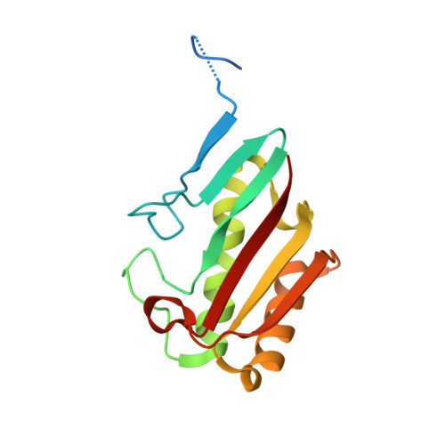
Crystal structures of the putative endoribonuclease L-PSP from Entamoeba histolytica.

Ojuromi, O.T.Giwa, A.O.Gardberg, A.Subramanian, S.Myler, P.J.Abendroth, J.Staker, B.Asojo, O.A.

(2025) Acta Crystallogr F Struct Biol Commun 81: 226-234

  • DOI: https://doi.org/10.1107/S2053230X25003875
  • Primary Citation Related Structures: 
    3M1X, 3M4S, 3MQW

  • PubMed Abstract: 

    Entamoeba histolytica causes amebiasis, a neglected disease that kills ∼100 000 people globally each year. Due to emerging drug resistance, E. histolytica is one of the target organisms for structure-based drug discovery by the Seattle Structural Genomics Center for Infectious Disease (SSGCID). Purification, crystallization and three structures of the putative drug target endoribonuclease L-PSP from E. histolytica (EhL-PSP) are presented. EhL-PSP has a two-layer α/β-sandwich with structural homology to endoribonuclease L-PSP. All three structures reveal the prototypical YjgF/YER057c/UK114 family trimer topology with accessible allosteric active sites. Citrate molecules from the crystallization solution are bound to the allosteric site in two of the three reported structures. The large allosteric site of EhL-PSP is well conserved with bacterial YjgF/YER057c/UK114 family members and could be targeted for inhibition, drug discovery or repurposing.


  • Organizational Affiliation
    • Zoology and Environmental Biology, Lagos State University, Nigeria.

Macromolecule Content 

  • Total Structure Weight: 96.28 kDa 
  • Atom Count: 6,943 
  • Modeled Residue Count: 817 
  • Deposited Residue Count: 888 
  • Unique protein chains: 1

Macromolecules

Find similar proteins by:|  3D Structure
Entity ID: 1
MoleculeChains  Sequence LengthOrganismDetailsImage
putative Endoribonuclease L-PSP
A, B, C, D, E
A, B, C, D, E, F
148Entamoeba histolyticaMutation(s): 0 
Gene Names: EHI_087570HM-1:IMSS
UniProt
Find proteins for C4LXT9 (Entamoeba histolytica (strain ATCC 30459 / HM-1:IMSS / ABRM))
Explore C4LXT9 
Go to UniProtKB:  C4LXT9
Entity Groups
Sequence Clusters30% Identity50% Identity70% Identity90% Identity95% Identity100% Identity
UniProt GroupC4LXT9
Sequence Annotations
Expand
Reference Sequence

Small Molecules

Ligands 3 Unique
IDChains Name / Formula / InChI Key2D Diagram3D Interactions
FLC

Query on FLC


Download:Ideal Coordinates CCD File
G [auth A],
I [auth B],
M [auth C],
Q [auth D],
U [auth E]
CITRATE ANION
C6 H5 O7
KRKNYBCHXYNGOX-UHFFFAOYSA-K
SO4

Query on SO4


Download:Ideal Coordinates CCD File
J [auth B]
K [auth B]
N [auth C]
O [auth C]
R [auth D]
J [auth B],
K [auth B],
N [auth C],
O [auth C],
R [auth D],
T [auth E],
W [auth F]
SULFATE ION
O4 S
QAOWNCQODCNURD-UHFFFAOYSA-L
GOL

Query on GOL


Download:Ideal Coordinates CCD File
H [auth A]
L [auth B]
P [auth C]
S [auth D]
V [auth E]
H [auth A],
L [auth B],
P [auth C],
S [auth D],
V [auth E],
X [auth F]
GLYCEROL
C3 H8 O3
PEDCQBHIVMGVHV-UHFFFAOYSA-N

Experimental Data & Validation

Experimental Data

  • Method: X-RAY DIFFRACTION
  • Resolution: 1.80 Å
  • R-Value Free:  0.183 (Depositor), 0.190 (DCC) 
  • R-Value Work:  0.147 (Depositor), 0.153 (DCC) 
  • R-Value Observed: 0.148 (Depositor) 
Space Group: P 21 21 21
Unit Cell:
Length ( Å )Angle ( ˚ )
a = 84α = 90
b = 99.72β = 90
c = 106.1γ = 90
Software Package:
Software NamePurpose
Blu-Icedata collection
PHASERphasing
REFMACrefinement
XDSdata reduction
XSCALEdata scaling

Structure Validation

View Full Validation Report


Entry History 

Deposition Data

Revision History  (Full details and data files)

  • Version 1.0: 2010-05-12
    Type: Initial release
  • Version 1.1: 2011-07-13
    Changes: Version format compliance
  • Version 1.2: 2023-09-06
    Changes: Data collection, Database references, Derived calculations, Refinement description
  • Version 1.3: 2025-12-24
    Changes: Database references, Structure summary