3MOJ | pdb_00003moj

Structure of the RNA binding domain of the Bacillus subtilis YxiN protein complexed with a fragment of 23S ribosomal RNA


Experimental Data Snapshot

  • Method: X-RAY DIFFRACTION
  • Resolution: 2.90 Å
  • R-Value Free: 
    0.275 (Depositor), 0.286 (DCC) 
  • R-Value Work: 
    0.209 (Depositor), 0.238 (DCC) 
  • R-Value Observed: 
    0.216 (Depositor) 

wwPDB Validation 3D Report Full Report

Validation slider image for 3MOJ

This is version 1.2 of the entry. See complete history

Literature

Structure of the RNA binding domain of a DEAD-box helicase bound to its ribosomal RNA target reveals a novel mode of recognition by an RNA recognition motif.

Hardin, J.W.Hu, Y.X.McKay, D.B.

(2010) J Mol Biology 402: 412-427

  • DOI: https://doi.org/10.1016/j.jmb.2010.07.040
  • Primary Citation Related Structures: 
    3MOJ

  • PubMed Abstract: 

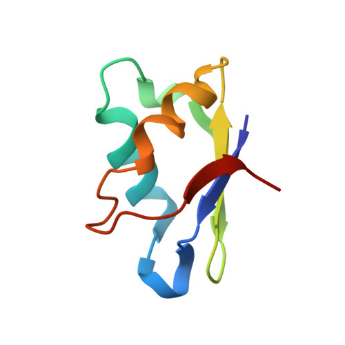
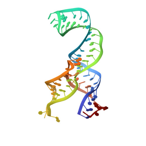
    DEAD-box RNA helicases of the bacterial DbpA subfamily are localized to their biological substrate when a carboxy-terminal RNA recognition motif domain binds tightly and specifically to a segment of 23S ribosomal RNA (rRNA) that includes hairpin 92 of the peptidyl transferase center. A complex between a fragment of 23S rRNA and the RNA binding domain (RBD) of the Bacillus subtilis DbpA protein YxiN was crystallized and its structure was determined to 2.9 A resolution, revealing an RNA recognition mode that differs from those observed with other RNA recognition motifs. The RBD is bound between two RNA strands at a three-way junction. Multiple phosphates of the RNA backbone interact with an electropositive band generated by lysines of the RBD. Nucleotides of the single-stranded loop of hairpin 92 interact with the RBD, including the guanosine base of G2553, which forms three hydrogen bonds with the peptide backbone. A G2553U mutation reduces the RNA binding affinity by 2 orders of magnitude, confirming that G2553 is a sequence specificity determinant in RNA binding. Binding of the RBD to 23S rRNA in the late stages of ribosome subunit maturation would position the ATP-binding duplex destabilization fragment of the protein for interaction with rRNA in the peptidyl transferase cleft of the subunit, allowing it to "melt out" unstable secondary structures and allow proper folding.


  • Organizational Affiliation
    • Department of Chemistry and Biochemistry, University of Colorado, Boulder, CO 80301, USA.

Macromolecule Content 

  • Total Structure Weight: 32.15 kDa 
  • Atom Count: 2,043 
  • Modeled Residue Count: 144 
  • Deposited Residue Count: 150 
  • Unique protein chains: 1
  • Unique nucleic acid chains: 1

Macromolecules


Find similar proteins by:|  3D Structure
Entity ID: 2
MoleculeChains  Sequence LengthOrganismDetailsImage
ATP-dependent RNA helicase dbpA76Bacillus subtilisMutation(s): 0 
Gene Names: dbpAdeaDyxiNBSU39110SS8E
EC: 3.6.1 (PDB Primary Data), 3.6.4.13 (UniProt)
UniProt
Find proteins for P42305 (Bacillus subtilis (strain 168))
Explore P42305 
Go to UniProtKB:  P42305
Entity Groups
Sequence Clusters30% Identity50% Identity70% Identity90% Identity95% Identity100% Identity
UniProt GroupP42305
Sequence Annotations
Expand
Reference Sequence
Find similar nucleic acids by:  (by identity cutoff) 
Entity ID: 1
MoleculeChains LengthOrganismImage
RNA (69-MER)74Escherichia coli
Sequence Annotations
Expand
Reference Sequence

Experimental Data & Validation

Experimental Data

  • Method: X-RAY DIFFRACTION
  • Resolution: 2.90 Å
  • R-Value Free:  0.275 (Depositor), 0.286 (DCC) 
  • R-Value Work:  0.209 (Depositor), 0.238 (DCC) 
  • R-Value Observed: 0.216 (Depositor) 
Space Group: P 21 21 21
Unit Cell:
Length ( Å )Angle ( ˚ )
a = 45.01α = 90
b = 91.63β = 90
c = 115.28γ = 90
Software Package:
Software NamePurpose
ALSdata collection
PHENIXmodel building
PHENIXrefinement
HKL-2000data reduction
HKL-2000data scaling
PHENIXphasing

Structure Validation

View Full Validation Report


Entry History 

Deposition Data

Revision History  (Full details and data files)

  • Version 1.0: 2010-08-04
    Type: Initial release
  • Version 1.1: 2011-07-13
    Changes: Version format compliance
  • Version 1.2: 2024-02-21
    Changes: Data collection, Database references