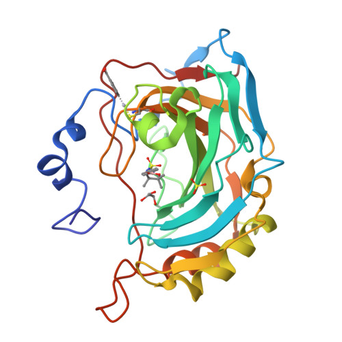
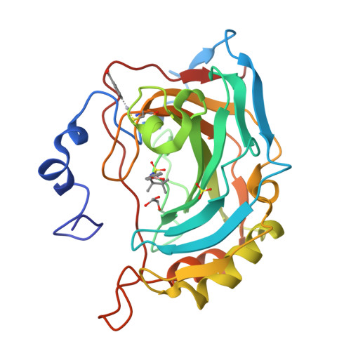
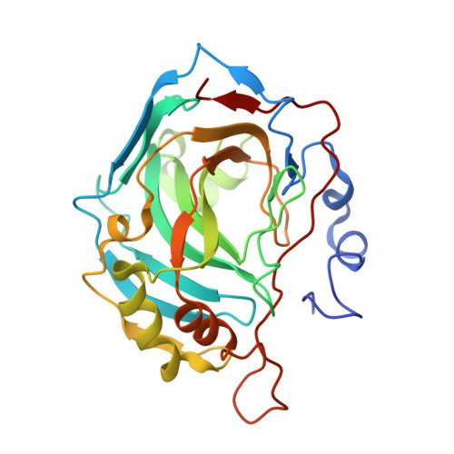
3MNU

Carbonic anhydrase inhibitors: crystallographic and solution binding studies for the interaction of a boron containing aromatic sulfamide with mammalian isoforms I-XV


Experimental Data Snapshot

  • Method: X-RAY DIFFRACTION
  • Resolution: 1.80 Å
  • R-Value Free: 0.205 
  • R-Value Work: 0.174 

Starting Model: experimental
View more details

wwPDB Validation   3D Report Full Report


Ligand Structure Quality Assessment 


This is version 1.2 of the entry. See complete history


Literature

Carbonic anhydrase inhibitors: crystallographic and solution binding studies for the interaction of a boron-containing aromatic sulfamide with mammalian isoforms I-XV.

Di Fiore, A.Monti, S.M.Innocenti, A.Winum, J.Y.De Simone, G.Supuran, C.T.

(2010) Bioorg Med Chem Lett 20: 3601-3605

  • DOI: https://doi.org/10.1016/j.bmcl.2010.04.114
  • Primary Citation of Related Structures:  
    3MNU

  • PubMed Abstract: 

    We investigated the inhibition of carbonic anhydrase (CA, EC 4.2.1.1) isoforms I-XV with 4-(4,4,5,5-tetramethyl-1,3,2-dioxaborolan-2-yl)phenylsulfamide and other simple or sugar sulfamides, a class of less investigated CA inhibitors (CAIs). The crystal structure of the adduct of hCA II with the boron-substituted sulfamide shows the organic scaffold of this compound bound in the hydrophilic half of the active site where it makes a large number of van der Waals contacts with Ile91, Gln92, Val121, Phe131, Leu198, and Thr200. The data here reported provide further insights into sulfamide binding mechanism confirming that this zinc-binding group could be usefully exploited for obtaining new potent and selective CAIs.


  • Organizational Affiliation

    Istituto di Biostrutture e Bioimmagini-CNR, Naples, Italy.


Macromolecules
Find similar proteins by:  (by identity cutoff)  |  3D Structure
Entity ID: 1
MoleculeChains Sequence LengthOrganismDetailsImage
Carbonic anhydrase 2259Homo sapiensMutation(s): 0 
EC: 4.2.1.1 (PDB Primary Data), 4.2.1.69 (UniProt)
UniProt & NIH Common Fund Data Resources
Find proteins for P00918 (Homo sapiens)
Explore P00918 
Go to UniProtKB:  P00918
PHAROS:  P00918
GTEx:  ENSG00000104267 
Entity Groups  
Sequence Clusters30% Identity50% Identity70% Identity90% Identity95% Identity100% Identity
UniProt GroupP00918
Sequence Annotations
Expand
  • Reference Sequence
Small Molecules
Ligands 5 Unique
IDChains Name / Formula / InChI Key2D Diagram3D Interactions
MBO
Query on MBO

Download Ideal Coordinates CCD File 
C [auth A]MERCURIBENZOIC ACID
C7 H5 Hg O2
FVFZSVRSDNUCGG-UHFFFAOYSA-N
BON
Query on BON

Download Ideal Coordinates CCD File 
E [auth A]N-[4-(4,4,5,5-tetramethyl-1,3,2-dioxaborolan-2-yl)phenyl]sulfamide
C12 H19 B N2 O4 S
YQWUJQMJCQBCRA-UHFFFAOYSA-N
SO4
Query on SO4

Download Ideal Coordinates CCD File 
F [auth A]SULFATE ION
O4 S
QAOWNCQODCNURD-UHFFFAOYSA-L
GOL
Query on GOL

Download Ideal Coordinates CCD File 
D [auth A]GLYCEROL
C3 H8 O3
PEDCQBHIVMGVHV-UHFFFAOYSA-N
ZN
Query on ZN

Download Ideal Coordinates CCD File 
B [auth A]ZINC ION
Zn
PTFCDOFLOPIGGS-UHFFFAOYSA-N
Binding Affinity Annotations 
IDSourceBinding Affinity
BON PDBBind:  3MNU Ki: 48 (nM) from 1 assay(s)
Experimental Data & Validation

Experimental Data

  • Method: X-RAY DIFFRACTION
  • Resolution: 1.80 Å
  • R-Value Free: 0.205 
  • R-Value Work: 0.174 
  • Space Group: P 1 21 1
Unit Cell:
Length ( Å )Angle ( ˚ )
a = 42.08α = 90
b = 41.33β = 104.45
c = 71.88γ = 90
Software Package:
Software NamePurpose
CNSrefinement
DENZOdata reduction
SCALEPACKdata scaling
CNSphasing

Structure Validation

View Full Validation Report


Ligand Structure Quality Assessment 


Entry History 

Deposition Data

Revision History  (Full details and data files)

  • Version 1.0: 2010-06-16
    Type: Initial release
  • Version 1.1: 2011-07-13
    Changes: Version format compliance
  • Version 1.2: 2023-09-06
    Changes: Data collection, Database references, Derived calculations, Refinement description