3MMC | pdb_00003mmc

Structure of the dissimilatory sulfite reductase from Archaeoglobus fulgidus


Experimental Data Snapshot

  • Method: X-RAY DIFFRACTION
  • Resolution: 2.04 Å
  • R-Value Free: 
    0.223 (Depositor), 0.255 (DCC) 
  • R-Value Work: 
    0.186 (Depositor), 0.227 (DCC) 
  • R-Value Observed: 
    0.188 (Depositor) 

wwPDB Validation 3D Report Full Report

Validation slider image for 3MMC

Ligand Structure Quality Assessment 


This is version 2.1 of the entry. See complete history

Literature

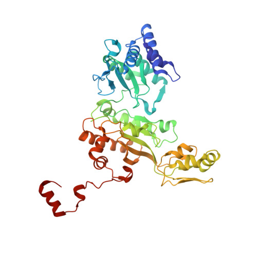
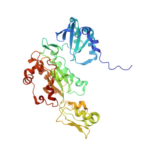
Structure of the dissimilatory sulfite reductase from the hyperthermophilic archaeon Archaeoglobus fulgidus.

Schiffer, A.Parey, K.Warkentin, E.Diederichs, K.Huber, H.Stetter, K.O.Kroneck, P.M.Ermler, U.

(2008) J Mol Biology 379: 1063-1074

  • DOI: https://doi.org/10.1016/j.jmb.2008.04.027
  • Primary Citation Related Structures: 
    3MMC

  • PubMed Abstract: 

    Conservation of energy based on the reduction of sulfate is of fundamental importance for the biogeochemical sulfur cycle. A key enzyme of this ancient anaerobic process is the dissimilatory sulfite reductase (dSir), which catalyzes the six-electron reduction of sulfite to hydrogen sulfide under participation of a unique magnetically coupled siroheme-[4Fe-4S] center. We determined the crystal structure of the enzyme from the sulfate-reducing archaeon Archaeoglobus fulgidus at 2-A resolution and compared it with that of the phylogenetically related assimilatory Sir (aSir). dSir is organized as a heterotetrameric (alphabeta)(2) complex composed of two catalytically independent alphabeta heterodimers. In contrast, aSir is a monomeric protein built of two fused modules that are structurally related to subunits alpha and beta except for a ferredoxin domain inserted only into the subunits of dSir. The [4Fe-4S] cluster of this ferredoxin domain is considered as the terminal redox site of the electron transfer pathway to the siroheme-[4Fe-4S] center in dSir. While aSir binds one siroheme-[4Fe-4S] center, dSir harbors two of them within each alphabeta heterodimer. Surprisingly, only one siroheme-[4Fe-4S] center in each alphabeta heterodimer is catalytically active, whereas access to the second one is blocked by a tryptophan residue. The spatial proximity of the functional and structural siroheme-[4Fe-4S] centers suggests that the catalytic activity at one active site was optimized during evolution at the expense of the enzymatic competence of the other. The sulfite binding mode and presumably the mechanism of sulfite reduction appear to be largely conserved between dSir and aSir. In addition, a scenario for the evolution of Sirs is proposed.


  • Organizational Affiliation
    • Fachbereich Biologie, Mathematisch-Naturwissenschaftliche Sektion, Universität Konstanz, D-78457 Konstanz, Germany.

Macromolecule Content 

  • Total Structure Weight: 185 kDa 
  • Atom Count: 13,324 
  • Modeled Residue Count: 1,560 
  • Deposited Residue Count: 1,568 
  • Unique protein chains: 2

Macromolecules

Find similar proteins by:|  3D Structure
Entity ID: 1
MoleculeChains  Sequence LengthOrganismDetailsImage
Sulfite reductase, dissimilatory-type subunit alphaA,
C [auth D]
418Archaeoglobus fulgidusMutation(s): 0 
EC: 1.8.99.3 (PDB Primary Data), 1.8.1.22 (UniProt)
UniProt
Find proteins for Q59109 (Archaeoglobus fulgidus (strain ATCC 49558 / DSM 4304 / JCM 9628 / NBRC 100126 / VC-16))
Explore Q59109 
Go to UniProtKB:  Q59109
Entity Groups
Sequence Clusters30% Identity50% Identity70% Identity90% Identity95% Identity100% Identity
UniProt GroupQ59109
Sequence Annotations
Expand
Reference Sequence
Find similar proteins by:|  3D Structure
Entity ID: 2
MoleculeChains  Sequence LengthOrganismDetailsImage
Sulfite reductase, dissimilatory-type subunit betaB,
D [auth E]
366Archaeoglobus fulgidusMutation(s): 0 
EC: 1.8.99.3 (PDB Primary Data), 1.8.1.22 (UniProt)
UniProt
Find proteins for Q59110 (Archaeoglobus fulgidus (strain ATCC 49558 / DSM 4304 / JCM 9628 / NBRC 100126 / VC-16))
Explore Q59110 
Go to UniProtKB:  Q59110
Entity Groups
Sequence Clusters30% Identity50% Identity70% Identity90% Identity95% Identity100% Identity
UniProt GroupQ59110
Sequence Annotations
Expand
Reference Sequence

Small Molecules

Ligands 3 Unique
IDChains Name / Formula / InChI Key2D Diagram3D Interactions
SRM

Query on SRM


Download:Ideal Coordinates CCD File
E [auth A],
K [auth B],
L [auth D],
Q [auth E]
SIROHEME
C42 H44 Fe N4 O16
DLKSSIHHLYNIKN-MWBYXLBFSA-L
SF4

Query on SF4


Download:Ideal Coordinates CCD File
F [auth A]
G [auth A]
I [auth B]
J [auth B]
M [auth D]
F [auth A],
G [auth A],
I [auth B],
J [auth B],
M [auth D],
N [auth D],
O [auth E],
P [auth E]
IRON/SULFUR CLUSTER
Fe4 S4
LJBDFODJNLIPKO-UHFFFAOYSA-N
GOL

Query on GOL


Download:Ideal Coordinates CCD File
H [auth A]GLYCEROL
C3 H8 O3
PEDCQBHIVMGVHV-UHFFFAOYSA-N

Experimental Data & Validation

Experimental Data

  • Method: X-RAY DIFFRACTION
  • Resolution: 2.04 Å
  • R-Value Free:  0.223 (Depositor), 0.255 (DCC) 
  • R-Value Work:  0.186 (Depositor), 0.227 (DCC) 
  • R-Value Observed: 0.188 (Depositor) 
Space Group: P 1 21 1
Unit Cell:
Length ( Å )Angle ( ˚ )
a = 94.91α = 90
b = 69.26β = 107
c = 147.21γ = 90
Software Package:
Software NamePurpose
HKL-2000data collection
SHARPphasing
REFMACrefinement
HKL-2000data reduction
XDSdata scaling

Structure Validation

View Full Validation Report


Ligand Structure Quality Assessment 


Entry History 

Deposition Data

Revision History  (Full details and data files)

  • Version 1.0: 2010-05-12
    Type: Initial release
  • Version 1.1: 2011-07-13
    Changes: Advisory, Refinement description, Version format compliance
  • Version 2.0: 2023-12-27
    Changes: Data collection, Database references, Derived calculations, Non-polymer description, Structure summary
  • Version 2.1: 2024-11-27
    Changes: Structure summary