3MLR | pdb_00003mlr

Crystal structure of anti-HIV-1 V3 Fab 2557 in complex with a NY5 V3 peptide


Experimental Data Snapshot

  • Method: X-RAY DIFFRACTION
  • Resolution: 1.80 Å
  • R-Value Free: 
    0.216 (Depositor), 0.216 (DCC) 
  • R-Value Work: 
    0.181 (Depositor), 0.181 (DCC) 

wwPDB Validation 3D Report Full Report

Validation slider image for 3MLR

This is version 1.3 of the entry. See complete history

Literature

Conserved structural elements in the V3 crown of HIV-1 gp120.

Jiang, X.Burke, V.Totrov, M.Williams, C.Cardozo, T.Gorny, M.K.Zolla-Pazner, S.Kong, X.P.

(2010) Nat Struct Mol Biol 17: 955-961

  • DOI: https://doi.org/10.1038/nsmb.1861
  • Primary Citation Related Structures: 
    3GO1, 3MLR, 3MLS, 3MLT, 3MLU, 3MLV, 3MLW, 3MLX, 3MLY, 3MLZ

  • PubMed Abstract: 

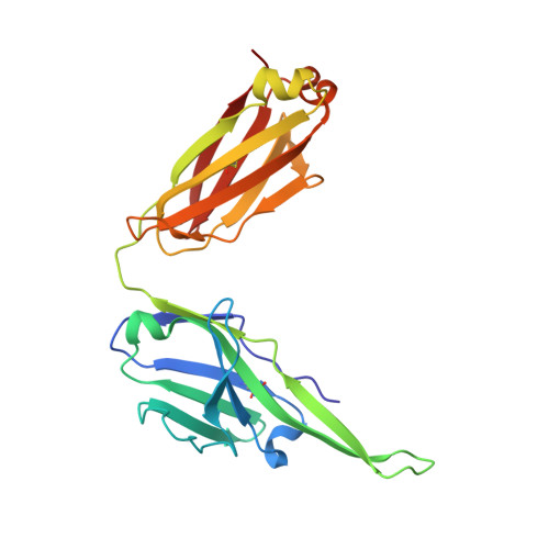
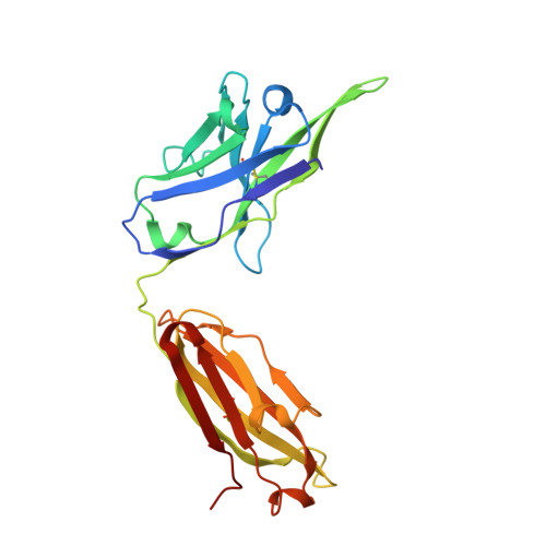
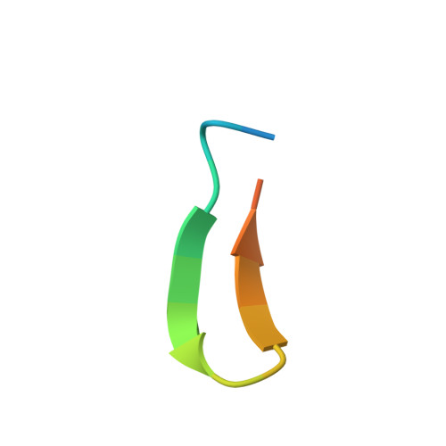
    Binding of the third variable region (V3) of the HIV-1 envelope glycoprotein gp120 to the cell-surface coreceptors CCR5 or CXCR4 during viral entry suggests that there are conserved structural elements in this sequence-variable region. These conserved elements could serve as epitopes to be targeted by a vaccine against HIV-1. Here we perform a systematic structural analysis of representative human anti-V3 monoclonal antibodies in complex with V3 peptides, revealing that the crown of V3 has four conserved structural elements: an arch, a band, a hydrophobic core and the peptide backbone. These are either unaffected by or are subject to minimal sequence variation. As these regions are targeted by cross-clade neutralizing human antibodies, they provide a blueprint for the design of vaccine immunogens that could elicit broadly cross-reactive protective antibodies.


  • Organizational Affiliation
    • Department of Biochemistry, New York University School of Medicine, New York, New York, USA.

Macromolecule Content 

  • Total Structure Weight: 49.72 kDa 
  • Atom Count: 3,927 
  • Modeled Residue Count: 459 
  • Deposited Residue Count: 465 
  • Unique protein chains: 3

Macromolecules

Find similar proteins by:|  3D Structure
Entity ID: 1
MoleculeChains  Sequence LengthOrganismDetailsImage
Human monoclonal anti-HIV-1 gp120 V3 antibody 2557 Fab light chainA [auth L]219Homo sapiensMutation(s): 0 
Find similar proteins by:|  3D Structure
Entity ID: 2
MoleculeChains  Sequence LengthOrganismDetailsImage
Human monoclonal anti-HIV-1 gp120 V3 antibody 2557 Fab heavy chainB [auth H]226Homo sapiensMutation(s): 0 
Find similar proteins by:  Sequence   |   3D Structure  
Entity ID: 3
MoleculeChains  Sequence LengthOrganismDetailsImage
HIV-1 gp120 third variable region (V3) crownC [auth P]20Human immunodeficiency virus type 1 (NEW YORK-5 ISOLATE)Mutation(s): 0 
UniProt
Find proteins for P12490 (Human immunodeficiency virus type 1 group M subtype B (isolate NY5))
Explore P12490 
Go to UniProtKB:  P12490
Entity Groups
Sequence Clusters30% Identity50% Identity70% Identity90% Identity95% Identity100% Identity
UniProt GroupP12490
Sequence Annotations
Expand
Reference Sequence

Experimental Data & Validation

Experimental Data

  • Method: X-RAY DIFFRACTION
  • Resolution: 1.80 Å
  • R-Value Free:  0.216 (Depositor), 0.216 (DCC) 
  • R-Value Work:  0.181 (Depositor), 0.181 (DCC) 
Space Group: P 1
Unit Cell:
Length ( Å )Angle ( ˚ )
a = 42.414α = 87.86
b = 43.076β = 85.5
c = 58.212γ = 85.82
Software Package:
Software NamePurpose
DENZOdata reduction
SCALEPACKdata scaling
CNSrefinement
PDB_EXTRACTdata extraction
ADSCdata collection
HKL-2000data reduction
MOLREPphasing

Structure Validation

View Full Validation Report


Entry History 

Deposition Data

  • Released Date: 2010-07-14 
  • Deposition Author(s): Kong, X.-P.

Revision History  (Full details and data files)

  • Version 1.0: 2010-07-14
    Type: Initial release
  • Version 1.1: 2011-07-13
    Changes: Version format compliance
  • Version 1.2: 2017-11-08
    Changes: Refinement description
  • Version 1.3: 2024-11-20
    Changes: Data collection, Database references, Structure summary