3MJV | pdb_00003mjv

Structure of A-type Ketoreductases from Modular Polyketide Synthase


Experimental Data Snapshot

  • Method: X-RAY DIFFRACTION
  • Resolution: 1.46 Å
  • R-Value Free: 
    0.236 (Depositor), 0.238 (DCC) 
  • R-Value Work: 
    0.199 (Depositor), 0.200 (DCC) 
  • R-Value Observed: 
    0.201 (Depositor) 

Starting Model: experimental
View more details

wwPDB Validation 3D Report Full Report

Validation slider image for 3MJV

Ligand Structure Quality Assessment 


This is version 1.3 of the entry. See complete history

Literature

Structural and Functional Analysis of A-Type Ketoreductases from the Amphotericin Modular Polyketide Synthase.

Zheng, J.Taylor, C.A.Piasecki, S.K.Keatinge-Clay, A.T.

(2010) Structure 18: 913-922

  • DOI: https://doi.org/10.1016/j.str.2010.04.015
  • Primary Citation Related Structures: 
    3MJC, 3MJE, 3MJS, 3MJT, 3MJV

  • PubMed Abstract: 

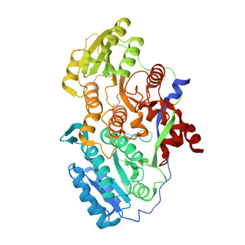
    Complex polyketides are characterized by multiple chiral centers harboring hydroxyl and alkyl substituents. To investigate the mechanisms by which these stereocenters are set, several high-resolution structures of the ketoreductase (KR) domain from the second module of the amphotericin modular polyketide synthase (PKS) were solved. This first structural analysis of an A-type KR helps reveal how these KRs direct polyketide intermediates into their active sites from the side opposite that used by B-type KRs, resulting in a beta-hydroxyl group of opposite stereochemistry. A comparison of structures obtained in the absence and presence of ligands reveals an induced fit mechanism that is important for catalysis. Activity assays of mutants of KRs from the first and second modules of the amphotericin PKS reveal the relative contributions of several active site residues toward catalysis and stereocontrol. Together, these results highlight the possibility of region-specific modification of polyketides through active site engineering of KRs.


  • Organizational Affiliation
    • Department of Chemistry and Biochemistry, The University of Texas at Austin, Austin, TX 78712-0165, USA.

Macromolecule Content 

  • Total Structure Weight: 104.26 kDa 
  • Atom Count: 7,547 
  • Modeled Residue Count: 951 
  • Deposited Residue Count: 992 
  • Unique protein chains: 1

Macromolecules

Find similar proteins by:|  3D Structure
Entity ID: 1
MoleculeChains  Sequence LengthOrganismDetailsImage
AmphB
A, B
496Streptomyces nodosusMutation(s): 1 
Gene Names: amphB
EC: 1.1.1.100
UniProt
Find proteins for Q93NW7 (Streptomyces nodosus)
Explore Q93NW7 
Go to UniProtKB:  Q93NW7
Entity Groups
Sequence Clusters30% Identity50% Identity70% Identity90% Identity95% Identity100% Identity
UniProt GroupQ93NW7
Sequence Annotations
Expand
Reference Sequence

Small Molecules

Ligands 1 Unique
IDChains Name / Formula / InChI Key2D Diagram3D Interactions
NDP

Query on NDP


Download:Ideal Coordinates CCD File
C [auth A],
D [auth B]
NADPH DIHYDRO-NICOTINAMIDE-ADENINE-DINUCLEOTIDE PHOSPHATE
C21 H30 N7 O17 P3
ACFIXJIJDZMPPO-NNYOXOHSSA-N

Experimental Data & Validation

Experimental Data

  • Method: X-RAY DIFFRACTION
  • Resolution: 1.46 Å
  • R-Value Free:  0.236 (Depositor), 0.238 (DCC) 
  • R-Value Work:  0.199 (Depositor), 0.200 (DCC) 
  • R-Value Observed: 0.201 (Depositor) 
Space Group: P 1
Unit Cell:
Length ( Å )Angle ( ˚ )
a = 61.502α = 72.93
b = 63.691β = 67.21
c = 71.92γ = 89.76
Software Package:
Software NamePurpose
ADSCdata collection
PHASESphasing
REFMACrefinement
HKL-2000data reduction
HKL-2000data scaling

Structure Validation

View Full Validation Report


Ligand Structure Quality Assessment 


Entry History 

Deposition Data

Revision History  (Full details and data files)

  • Version 1.0: 2010-08-18
    Type: Initial release
  • Version 1.1: 2011-07-13
    Changes: Version format compliance
  • Version 1.2: 2021-10-06
    Changes: Database references, Derived calculations
  • Version 1.3: 2023-09-06
    Changes: Data collection, Refinement description