3MJ6 | pdb_00003mj6

Crystal structure of the gammadelta T cell costimulatory receptor Junctional Adhesion Molecule-Like Protein, JAML


Experimental Data Snapshot

  • Method: X-RAY DIFFRACTION
  • Resolution: 2.19 Å
  • R-Value Free: 
    0.213 (Depositor), 0.216 (DCC) 
  • R-Value Work: 
    0.170 (Depositor), 0.174 (DCC) 
  • R-Value Observed: 
    0.172 (Depositor) 

Starting Model: experimental
View more details

wwPDB Validation 3D Report Full Report

Validation slider image for 3MJ6

Ligand Structure Quality Assessment 


This is version 2.2 of the entry. See complete history

Literature

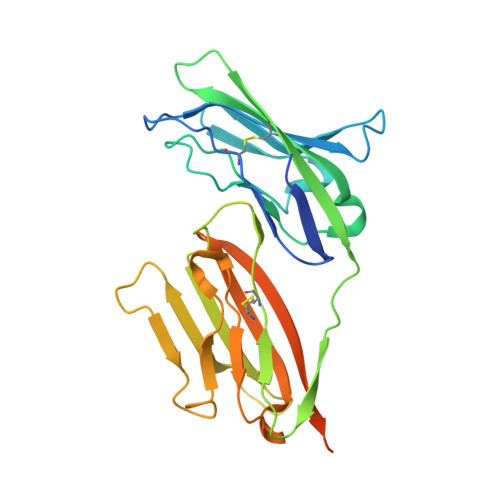
The molecular interaction of CAR and JAML recruits the central cell signal transducer PI3K.

Verdino, P.Witherden, D.A.Havran, W.L.Wilson, I.A.

(2010) Science 329: 1210-1214

  • DOI: https://doi.org/10.1126/science.1187996
  • Primary Citation Related Structures: 
    3MJ6, 3MJ7

  • PubMed Abstract: 

    Coxsackie and adenovirus receptor (CAR) is the primary cellular receptor for group B coxsackieviruses and most adenovirus serotypes and plays a crucial role in adenoviral gene therapy. Recent discovery of the interaction between junctional adhesion molecule-like protein (JAML) and CAR uncovered important functional roles in immunity, inflammation, and tissue homeostasis. Crystal structures of JAML ectodomain (2.2 angstroms) and its complex with CAR (2.8 angstroms) reveal an unusual immunoglobulin-domain assembly for JAML and a charged interface that confers high specificity. Biochemical and mutagenesis studies illustrate how CAR-mediated clustering of JAML recruits phosphoinositide 3-kinase (P13K) to a JAML intracellular sequence motif as delineated for the alphabeta T cell costimulatory receptor CD28. Thus, CAR and JAML are cell signaling receptors of the immune system with implications for asthma, cancer, and chronic nonhealing wounds.


  • Organizational Affiliation
    • Department of Molecular Biology, The Scripps Research Institute, 10550 North Torrey Pines Road, La Jolla, CA 92037, USA.

Macromolecule Content 

  • Total Structure Weight: 31.85 kDa 
  • Atom Count: 2,062 
  • Modeled Residue Count: 227 
  • Deposited Residue Count: 268 
  • Unique protein chains: 1

Macromolecules

Find similar proteins by:|  3D Structure
Entity ID: 1
MoleculeChains  Sequence LengthOrganismDetailsImage
Junctional adhesion molecule-like268Mus musculusMutation(s): 2 
Gene Names: Amica1Gm638Jaml
UniProt
Find proteins for Q80UL9 (Mus musculus)
Explore Q80UL9 
Go to UniProtKB:  Q80UL9
Entity Groups
Sequence Clusters30% Identity50% Identity70% Identity90% Identity95% Identity100% Identity
UniProt GroupQ80UL9
Glycosylation
Glycosylation Sites: 3Go to GlyGen: Q80UL9-1
Sequence Annotations
Expand
Reference Sequence

Oligosaccharides

Help  
Entity ID: 2
MoleculeChains Length2D Diagram GlycosylationD Interactions
alpha-L-fucopyranose-(1-6)-2-acetamido-2-deoxy-beta-D-glucopyranose
B
2N-Glycosylation
Glycosylation Resources
GlyTouCan: G86851RC
GlyCosmos: G86851RC
GlyGen: G86851RC

Small Molecules

Ligands 3 Unique
IDChains Name / Formula / InChI Key2D Diagram3D Interactions
NAG

Query on NAG


Download:Ideal Coordinates CCD File
C [auth A],
D [auth A]
2-acetamido-2-deoxy-beta-D-glucopyranose
C8 H15 N O6
OVRNDRQMDRJTHS-FMDGEEDCSA-N
FMT

Query on FMT


Download:Ideal Coordinates CCD File
F [auth A]
G [auth A]
H [auth A]
I [auth A]
J [auth A]
F [auth A],
G [auth A],
H [auth A],
I [auth A],
J [auth A],
K [auth A],
L [auth A],
M [auth A]
FORMIC ACID
C H2 O2
BDAGIHXWWSANSR-UHFFFAOYSA-N
NA

Query on NA


Download:Ideal Coordinates CCD File
E [auth A]SODIUM ION
Na
FKNQFGJONOIPTF-UHFFFAOYSA-N

Experimental Data & Validation

Experimental Data

  • Method: X-RAY DIFFRACTION
  • Resolution: 2.19 Å
  • R-Value Free:  0.213 (Depositor), 0.216 (DCC) 
  • R-Value Work:  0.170 (Depositor), 0.174 (DCC) 
  • R-Value Observed: 0.172 (Depositor) 
Space Group: P 43
Unit Cell:
Length ( Å )Angle ( ˚ )
a = 61.946α = 90
b = 61.946β = 90
c = 82.477γ = 90
Software Package:
Software NamePurpose
SHELXmodel building
RESOLVEmodel building
PHASERphasing
REFMACrefinement
HKL-2000data reduction
HKL-2000data scaling
SHELXphasing
RESOLVEphasing

Structure Validation

View Full Validation Report


Ligand Structure Quality Assessment 


Entry History 

Deposition Data

Revision History  (Full details and data files)

  • Version 1.0: 2010-09-22
    Type: Initial release
  • Version 1.1: 2011-07-13
    Changes: Version format compliance
  • Version 2.0: 2020-07-29
    Type: Remediation
    Reason: Carbohydrate remediation
    Changes: Atomic model, Data collection, Database references, Derived calculations, Structure summary
  • Version 2.1: 2024-04-03
    Changes: Data collection, Database references, Refinement description, Structure summary
  • Version 2.2: 2024-10-09
    Changes: Structure summary