3MGE | pdb_00003mge

X-ray Structure of Hexameric HIV-1 CA


Experimental Data Snapshot

  • Method: X-RAY DIFFRACTION
  • Resolution: 1.90 Å
  • R-Value Free: 
    0.255 (Depositor), 0.259 (DCC) 
  • R-Value Work: 
    0.230 (Depositor), 0.227 (DCC) 
  • R-Value Observed: 
    0.231 (Depositor) 

Starting Model: experimental
View more details

wwPDB Validation 3D Report Full Report

Validation slider image for 3MGE

This is version 1.4 of the entry. See complete history

Literature

Disulfide Bond Stabilization of the Hexameric Capsomer of Human Immunodeficiency Virus.

Pornillos, O.Ganser-Pornillos, B.K.Banumathi, S.Hua, Y.Yeager, M.

(2010) J Mol Biology 401: 985-995

  • DOI: https://doi.org/10.1016/j.jmb.2010.06.042
  • Primary Citation Related Structures: 
    3MGE

  • PubMed Abstract: 

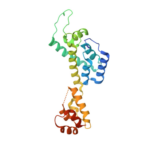
    The human immunodeficiency virus type 1 capsid is modeled as a fullerene cone that is composed of approximately 250 hexamers and 12 pentamers of the viral CA protein. Structures of CA hexamers have been difficult to obtain because the hexamer-stabilizing interactions are inherently weak, and CA tends to spontaneously assemble into capsid-like particles. Here, we describe a two-step biochemical strategy to obtain soluble CA hexamers for crystallization. First, the hexamer was stabilized by engineering disulfide cross-links (either A14C/E45C or A42C/T54C) between the N-terminal domains of adjacent subunits. Second, the cross-linked hexamers were prevented from polymerizing further into hyperstable capsid-like structures by mutations (W184A and M185A) that interfered with dimeric association between the C-terminal domains that link adjacent hexamers. The structures of two different cross-linked CA hexamers were nearly identical, and we combined the non-mutated portions of the structures to generate an atomic resolution model for the native hexamer. This hybrid approach for structure determination should be applicable to other viral capsomers and protein-protein complexes in general.


  • Organizational Affiliation
    • Department of Molecular Physiology and Biological Physics, University of Virginia, Charlottesville, VA 22908, USA.

Macromolecule Content 

  • Total Structure Weight: 25.55 kDa 
  • Atom Count: 1,747 
  • Modeled Residue Count: 204 
  • Deposited Residue Count: 231 
  • Unique protein chains: 1

Macromolecules

Find similar proteins by:|  3D Structure
Entity ID: 1
MoleculeChains  Sequence LengthOrganismDetailsImage
Capsid protein p24231Human immunodeficiency virus type 1 (NEW YORK-5 ISOLATE)Mutation(s): 4 
Gene Names: CAgag-pol
UniProt
Find proteins for P12497 (Human immunodeficiency virus type 1 group M subtype B (isolate NY5))
Explore P12497 
Go to UniProtKB:  P12497
Entity Groups
Sequence Clusters30% Identity50% Identity70% Identity90% Identity95% Identity100% Identity
UniProt GroupP12497
Sequence Annotations
Expand
Reference Sequence

Small Molecules

Ligands 1 Unique
IDChains Name / Formula / InChI Key2D Diagram3D Interactions
EDO

Query on EDO


Download:Ideal Coordinates CCD File
B [auth A]1,2-ETHANEDIOL
C2 H6 O2
LYCAIKOWRPUZTN-UHFFFAOYSA-N

Experimental Data & Validation

Experimental Data

  • Method: X-RAY DIFFRACTION
  • Resolution: 1.90 Å
  • R-Value Free:  0.255 (Depositor), 0.259 (DCC) 
  • R-Value Work:  0.230 (Depositor), 0.227 (DCC) 
  • R-Value Observed: 0.231 (Depositor) 
Space Group: P 6
Unit Cell:
Length ( Å )Angle ( ˚ )
a = 90.313α = 90
b = 90.313β = 90
c = 56.527γ = 120
Software Package:
Software NamePurpose
MOLREPphasing
PHENIXrefinement
HKL-2000data reduction
HKL-2000data scaling

Structure Validation

View Full Validation Report


Entry History 

Deposition Data

Revision History  (Full details and data files)

  • Version 1.0: 2010-07-21
    Type: Initial release
  • Version 1.1: 2011-07-13
    Changes: Version format compliance
  • Version 1.2: 2021-10-06
    Changes: Database references, Derived calculations
  • Version 1.3: 2023-09-06
    Changes: Data collection, Refinement description
  • Version 1.4: 2024-10-09
    Changes: Structure summary