3MFF | pdb_00003mff

1F1E8hu TCR


Experimental Data Snapshot

  • Method: X-RAY DIFFRACTION
  • Resolution: 2.00 Å
  • R-Value Free: 
    0.243 (Depositor), 0.244 (DCC) 
  • R-Value Work: 
    0.200 (Depositor), 0.248 (DCC) 
  • R-Value Observed: 
    0.202 (Depositor) 

wwPDB Validation 3D Report Full Report

Validation slider image for 3MFF

Ligand Structure Quality Assessment 


This is version 1.3 of the entry. See complete history

Literature

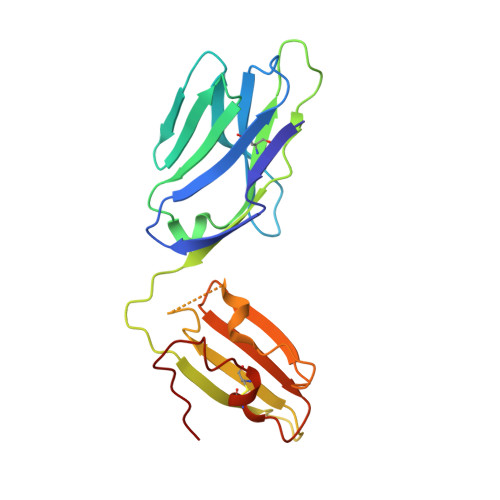
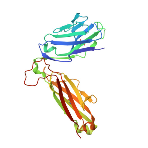
An alternative conformation of the T-cell receptor alpha constant region

van Boxel, G.I.Holmes, S.Fugger, L.Jones, E.Y.

(2010) J Mol Biology 400: 828-837

  • DOI: https://doi.org/10.1016/j.jmb.2010.05.053
  • Primary Citation Related Structures: 
    3MFF

  • PubMed Abstract: 

    Alphabeta T-cell receptors (TcRs) play a central role in cellular immune response. They are members of the Ig superfamily, with extracellular regions of the alpha and beta chains each comprising a V-type domain and a C-type domain. We have determined the ectodomain structure of an alphabeta TcR, which recognizes the autoantigen myelin basic protein. The 2.0-A-resolution structure reveals canonical main-chain conformations for the V(alpha), V(beta), and C(beta) domains, but the C(alpha) domain exhibits a main-chain conformation remarkably different from those previously reported for TcR crystal structures. The global IgC-like fold is maintained, but a piston-like rearrangement between BC and DE beta-turns results in beta-strand slippage. This substantial conformational change may represent a signaling intermediate. Our structure is the first example for the Ig fold of the increasingly recognized concept of "metamorphic proteins."


  • Organizational Affiliation
    • Division of Structural Biology, Wellcome Trust Centre for Human Genetics, The University of Oxford, Roosevelt Drive, Oxford OX3 7BN, UK.

Macromolecule Content 

  • Total Structure Weight: 100.55 kDa 
  • Atom Count: 7,784 
  • Modeled Residue Count: 873 
  • Deposited Residue Count: 884 
  • Unique protein chains: 2

Macromolecules

Find similar proteins by:|  3D Structure
Entity ID: 1
MoleculeChains  Sequence LengthOrganismDetailsImage
T cell receptor alpha chain
A, C
200Homo sapiensMutation(s): 0 
Find similar proteins by:|  3D Structure
Entity ID: 2
MoleculeChains  Sequence LengthOrganismDetailsImage
T cell receptor beta chain
B, D
242Homo sapiensMutation(s): 0 

Experimental Data & Validation

Experimental Data

  • Method: X-RAY DIFFRACTION
  • Resolution: 2.00 Å
  • R-Value Free:  0.243 (Depositor), 0.244 (DCC) 
  • R-Value Work:  0.200 (Depositor), 0.248 (DCC) 
  • R-Value Observed: 0.202 (Depositor) 
Space Group: P 1 21 1
Unit Cell:
Length ( Å )Angle ( ˚ )
a = 87.323α = 90
b = 74.016β = 91.59
c = 94.267γ = 90
Software Package:
Software NamePurpose
HKL-2000data collection
AMoREphasing
REFMACrefinement
HKL-2000data reduction
HKL-2000data scaling

Structure Validation

View Full Validation Report


Ligand Structure Quality Assessment 


Entry History 

Deposition Data

Revision History  (Full details and data files)

  • Version 1.0: 2010-06-02
    Type: Initial release
  • Version 1.1: 2011-07-13
    Changes: Version format compliance
  • Version 1.2: 2014-03-05
    Changes: Database references
  • Version 1.3: 2024-10-30
    Changes: Data collection, Database references, Derived calculations, Refinement description, Structure summary