3MC0 | pdb_00003mc0

Crystal Structure of Staphylococcal Enterotoxin G (SEG) in Complex with a Mouse T-cell Receptor beta Chain


Experimental Data Snapshot

  • Method: X-RAY DIFFRACTION
  • Resolution: 2.00 Å
  • R-Value Free: 
    0.211 (Depositor), 0.206 (DCC) 
  • R-Value Work: 
    0.179 (Depositor), 0.176 (DCC) 
  • R-Value Observed: 
    0.180 (Depositor) 

Starting Model: experimental
View more details

wwPDB Validation 3D Report Full Report

Validation slider image for 3MC0

This is version 1.3 of the entry. See complete history

Literature

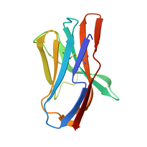
Crystal structure of staphylococcal enterotoxin G (SEG) in complex with a mouse T-cell receptor {beta} chain.

Fernandez, M.M.Cho, S.De Marzi, M.C.Kerzic, M.C.Robinson, H.Mariuzza, R.A.Malchiodi, E.L.

(2011) J Biological Chem 286: 1189-1195

  • DOI: https://doi.org/10.1074/jbc.M110.142471
  • Primary Citation Related Structures: 
    3MC0, 3OWE

  • PubMed Abstract: 

    Superantigens (SAgs) are bacterial or viral toxins that bind MHC class II (MHC-II) molecules and T-cell receptor (TCR) in a nonconventional manner, inducing T-cell activation that leads to inflammatory cytokine production, which may result in acute toxic shock. In addition, the emerging threat of purpura fulminans and community-associated meticillin-resistant Staphylococcus aureus emphasizes the importance of a better characterization of SAg binding to their natural ligands that may allow the development of reagents to neutralize their action. The three-dimensional structure of the complex between a mouse TCR β chain (mVβ8.2) and staphylococcal enterotoxin G (SEG) at 2.0 Å resolution revealed a binding site that does not conserve the "hot spots" present in mVβ8.2-SEC2, mVβ8.2-SEC3, mVβ8.2-SEB, and mVβ8.2-SPEA complexes. Analysis of the mVβ8.2-SEG interface allowed us to explain the higher affinity of this complex compared with the others, which may account for the early activation of T-cells bearing mVβ8.2 by SEG. This mode of interaction between SEG and mVβ8.2 could be an adaptive advantage to bestow on the pathogen a faster rate of colonization of the host.


  • Organizational Affiliation
    • Cátedra de Inmunología and Instituto de Estudios de la Inmunidad Humoral Prof Ricardo A. Margni, Consejo Nacional de Investigaciones Científicas y Técnicas, Facultad de Farmacia y Bioquímica, Universidad de Buenos Aires, Junín 956 4to P, 1113 Buenos Aires, Argentina.

Macromolecule Content 

  • Total Structure Weight: 79.7 kDa 
  • Atom Count: 5,841 
  • Modeled Residue Count: 673 
  • Deposited Residue Count: 696 
  • Unique protein chains: 2

Macromolecules

Find similar proteins by:|  3D Structure
Entity ID: 1
MoleculeChains  Sequence LengthOrganismDetailsImage
variable beta 8.2 mouse T cell receptor
A, C
109Mus musculusMutation(s): 8 
UniProt
Find proteins for A2NTY6 (Mus musculus)
Explore A2NTY6 
Go to UniProtKB:  A2NTY6
Entity Groups
Sequence Clusters30% Identity50% Identity70% Identity90% Identity95% Identity100% Identity
UniProt GroupA2NTY6
Sequence Annotations
Expand
Reference Sequence
Find similar proteins by:|  3D Structure
Entity ID: 2
MoleculeChains  Sequence LengthOrganismDetailsImage
Enterotoxin SEG
B, D
239Staphylococcus aureusMutation(s): 0 
UniProt
Find proteins for D0EMB6 (Staphylococcus aureus)
Explore D0EMB6 
Go to UniProtKB:  D0EMB6
Entity Groups
Sequence Clusters30% Identity50% Identity70% Identity90% Identity95% Identity100% Identity
UniProt GroupD0EMB6
Sequence Annotations
Expand
Reference Sequence

Experimental Data & Validation

Experimental Data

  • Method: X-RAY DIFFRACTION
  • Resolution: 2.00 Å
  • R-Value Free:  0.211 (Depositor), 0.206 (DCC) 
  • R-Value Work:  0.179 (Depositor), 0.176 (DCC) 
  • R-Value Observed: 0.180 (Depositor) 
Space Group: P 31 2 1
Unit Cell:
Length ( Å )Angle ( ˚ )
a = 91.185α = 90
b = 91.185β = 90
c = 233.401γ = 120
Software Package:
Software NamePurpose
ADSCdata collection
MOLREPphasing
REFMACrefinement
HKL-2000data reduction
HKL-2000data scaling

Structure Validation

View Full Validation Report


Entry History 

Deposition Data

Revision History  (Full details and data files)

  • Version 1.0: 2010-10-13
    Type: Initial release
  • Version 1.1: 2011-07-13
    Changes: Version format compliance
  • Version 1.2: 2023-09-06
    Changes: Data collection, Database references, Derived calculations, Refinement description
  • Version 1.3: 2024-10-16
    Changes: Structure summary