3M9Z | pdb_00003m9z

Crystal Structure of extracellular domain of mouse NKR-P1A


Experimental Data Snapshot

  • Method: X-RAY DIFFRACTION
  • Resolution: 1.70 Å
  • R-Value Free: 
    0.239 (Depositor), 0.218 (DCC) 
  • R-Value Work: 
    0.195 (Depositor), 0.201 (DCC) 
  • R-Value Observed: 
    0.197 (Depositor) 

Starting Model: experimental
View more details

wwPDB Validation 3D Report Full Report

Validation slider image for 3M9Z

This is version 1.4 of the entry. See complete history

Literature

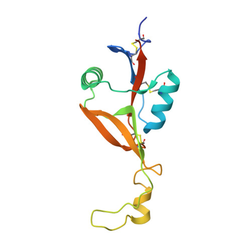
Molecular architecture of mouse activating NKR-P1 receptors.

Kolenko, P.Rozbesky, D.Vanek, O.Kopecky, V.Hofbauerova, K.Novak, P.Pompach, P.Hasek, J.Skalova, T.Bezouska, K.Dohnalek, J.

(2011) J Struct Biol 175: 434-441

  • DOI: https://doi.org/10.1016/j.jsb.2011.05.001
  • Primary Citation Related Structures: 
    3M9Z

  • PubMed Abstract: 

    Receptors belonging to NKR-P1 family and their specific Clr ligands form an alternative missing self recognition system critical in immunity against tumors and viruses, elimination of tumor cells subjected to genotoxic stress, activation of T cell dependent immune response, and hypertension. The three-dimensional structure of the extracellular domain of the mouse natural killer (NK) cell receptor mNKR-P1Aex has been determined by X-ray diffraction. The core of the C-type lectin domain (CTLD) is homologous to the other CTLD receptors whereas one quarter of the domain forms an extended loop interacting tightly with a neighboring loop in the crystal. This domain swapping mechanism results in a compact interaction interface. A second dimerization interface resembles the known arrangement of other CTLD NK receptors. A functional dimeric form of the receptor is suggested, with the loop, evolutionarily conserved within this family, proposed to participate in interactions with ligands.


  • Organizational Affiliation
    • Institute of Macromolecular Chemistry, v.v.i., Academy of Sciences of the Czech Republic, Heyrovského náměstí 2, 16206 Prague 6, Czech Republic.

Macromolecule Content 

  • Total Structure Weight: 16.1 kDa 
  • Atom Count: 1,134 
  • Modeled Residue Count: 124 
  • Deposited Residue Count: 139 
  • Unique protein chains: 1

Macromolecules

Find similar proteins by:|  3D Structure
Entity ID: 1
MoleculeChains  Sequence LengthOrganismDetailsImage
Killer cell lectin-like receptor subfamily B member 1A139Mus musculusMutation(s): 2 
Gene Names: Klrb1aLy55Ly55aNkrp1a
UniProt
Find proteins for P27811 (Mus musculus)
Explore P27811 
Go to UniProtKB:  P27811
Entity Groups
Sequence Clusters30% Identity50% Identity70% Identity90% Identity95% Identity100% Identity
UniProt GroupP27811
Sequence Annotations
Expand
Reference Sequence

Small Molecules

Ligands 1 Unique
IDChains Name / Formula / InChI Key2D Diagram3D Interactions
PO4

Query on PO4


Download:Ideal Coordinates CCD File
B [auth A]PHOSPHATE ION
O4 P
NBIIXXVUZAFLBC-UHFFFAOYSA-K

Experimental Data & Validation

Experimental Data

  • Method: X-RAY DIFFRACTION
  • Resolution: 1.70 Å
  • R-Value Free:  0.239 (Depositor), 0.218 (DCC) 
  • R-Value Work:  0.195 (Depositor), 0.201 (DCC) 
  • R-Value Observed: 0.197 (Depositor) 
Space Group: I 41 2 2
Unit Cell:
Length ( Å )Angle ( ˚ )
a = 59.912α = 90
b = 59.912β = 90
c = 159.303γ = 90
Software Package:
Software NamePurpose
MxCuBEdata collection
BALBESphasing
REFMACrefinement
MOSFLMdata reduction
SCALAdata scaling

Structure Validation

View Full Validation Report


Entry History 

Deposition Data

Revision History  (Full details and data files)

  • Version 1.0: 2011-04-06
    Type: Initial release
  • Version 1.1: 2011-07-13
    Changes: Version format compliance
  • Version 1.2: 2015-02-18
    Changes: Database references
  • Version 1.3: 2023-09-06
    Changes: Data collection, Database references, Derived calculations, Refinement description
  • Version 1.4: 2024-11-27
    Changes: Structure summary