3M9F | pdb_00003m9f

HIV protease complexed with compound 10b


Experimental Data Snapshot

  • Method: X-RAY DIFFRACTION
  • Resolution: 1.80 Å
  • R-Value Free: 
    0.230 (Depositor), 0.217 (DCC) 
  • R-Value Work: 
    0.187 (Depositor) 
  • R-Value Observed: 
    0.190 (Depositor) 

wwPDB Validation 3D Report Full Report

Validation slider image for 3M9F

Ligand Structure Quality Assessment 


This is version 1.3 of the entry. See complete history

Literature

Epsilon substituted lysinol derivatives as HIV-1 protease inhibitors.

Jones, K.L.Holloway, M.K.Su, H.P.Carroll, S.S.Burlein, C.Touch, S.DiStefano, D.J.Sanchez, R.I.Williams, T.M.Vacca, J.P.Coburn, C.A.

(2010) Bioorg Med Chem Lett 20: 4065-4068

  • DOI: https://doi.org/10.1016/j.bmcl.2010.05.082
  • Primary Citation Related Structures: 
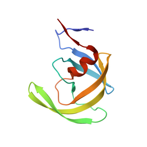
    3M9F

  • PubMed Abstract: 

    A series of HIV-1 protease inhibitors containing an epsilon substituted lysinol backbone was synthesized. Two novel synthetic routes using N-boc-L-glutamic acid alpha-benzyl ester and 2,6-diaminopimelic acid were developed. Incorporation of this epsilon substituent enabled access to the S2 pocket of the enzyme, affording high potency inhibitors. Modeling studies and synthetic efforts suggest the potency increase is due to both conformational bias and van der Waals interactions with the S2 pocket.


  • Organizational Affiliation
    • Department of Medicinal Chemistry, Merck Research Laboratories, PO Box 4, West Point, PA 19486, USA. kristen_jones@merck.com

Macromolecule Content 

  • Total Structure Weight: 23.07 kDa 
  • Atom Count: 1,730 
  • Modeled Residue Count: 198 
  • Deposited Residue Count: 198 
  • Unique protein chains: 1

Macromolecules

Find similar proteins by:|  3D Structure
Entity ID: 1
MoleculeChains  Sequence LengthOrganismDetailsImage
HIV-1 protease
A, B
99Human immunodeficiency virus 1Mutation(s): 0 
UniProt
Find proteins for Q9WFL7 (Human immunodeficiency virus type 1)
Explore Q9WFL7 
Go to UniProtKB:  Q9WFL7
Entity Groups
Sequence Clusters30% Identity50% Identity70% Identity90% Identity95% Identity100% Identity
UniProt GroupQ9WFL7
Sequence Annotations
Expand
Reference Sequence

Experimental Data & Validation

Experimental Data

  • Method: X-RAY DIFFRACTION
  • Resolution: 1.80 Å
  • R-Value Free:  0.230 (Depositor), 0.217 (DCC) 
  • R-Value Work:  0.187 (Depositor) 
  • R-Value Observed: 0.190 (Depositor) 
Space Group: P 21 21 2
Unit Cell:
Length ( Å )Angle ( ˚ )
a = 58.173α = 90
b = 85.934β = 90
c = 46.134γ = 90
Software Package:
Software NamePurpose
DENZOdata reduction
SCALEPACKdata scaling
REFMACrefinement
PDB_EXTRACTdata extraction
REFMACphasing

Structure Validation

View Full Validation Report


Ligand Structure Quality Assessment 


Entry History 

Deposition Data

  • Released Date: 2010-06-30 
  • Deposition Author(s): Su, H.P.

Revision History  (Full details and data files)

  • Version 1.0: 2010-06-30
    Type: Initial release
  • Version 1.1: 2011-07-13
    Changes: Version format compliance
  • Version 1.2: 2017-11-08
    Changes: Refinement description
  • Version 1.3: 2024-02-21
    Changes: Data collection, Database references, Derived calculations