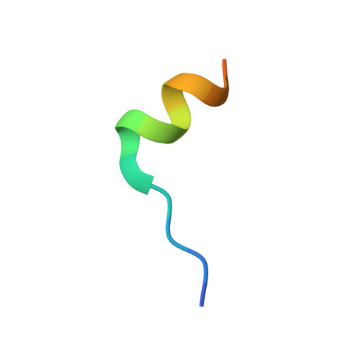
3M93

Complex crystal structure of Ascaris suum eIF4E-3 with m7G cap


Experimental Data Snapshot

  • Method: X-RAY DIFFRACTION
  • Resolution: 2.90 Å
  • R-Value Free: 0.294 
  • R-Value Work: 0.239 
  • R-Value Observed: 0.245 

Starting Model: experimental
View more details

wwPDB Validation   3D Report Full Report


Ligand Structure Quality Assessment 


This is version 2.0 of the entry. See complete history


Literature

Structural basis for nematode eIF4E binding an m2,2,7G-Cap and its implications for translation initiation.

Liu, W.Jankowska-Anyszka, M.Piecyk, K.Dickson, L.Wallace, A.Niedzwiecka, A.Stepinski, J.Stolarski, R.Darzynkiewicz, E.Kieft, J.Zhao, R.Jones, D.N.Davis, R.E.

(2011) Nucleic Acids Res 39: 8820-8832

  • DOI: https://doi.org/10.1093/nar/gkr650
  • Primary Citation of Related Structures:  
    3M93, 3M94

  • PubMed Abstract: 

    Metazoan spliced leader (SL) trans-splicing generates mRNAs with an m(2,2,7)G-cap and a common downstream SL RNA sequence. The mechanism for eIF4E binding an m²²⁷G-cap is unknown. Here, we describe the first structure of an eIF4E with an m(2,2,7)G-cap and compare it to the cognate m⁷G-eIF4E complex. These structures and Nuclear Magnetic Resonance (NMR) data indicate that the nematode Ascaris suum eIF4E binds the two different caps in a similar manner except for the loss of a single hydrogen bond on binding the m(2,2,7)G-cap. Nematode and mammalian eIF4E both have a low affinity for m(2,2,7)G-cap compared with the m⁷G-cap. Nematode eIF4E binding to the m⁷G-cap, m(2,2,7)G-cap and the m(2,2,7)G-SL 22-nt RNA leads to distinct eIF4E conformational changes. Additional interactions occur between Ascaris eIF4E and the SL on binding the m(2,2,7)G-SL. We propose interactions between Ascaris eIF4E and the SL impact eIF4G and contribute to translation initiation, whereas these interactions do not occur when only the m(2,2,7)G-cap is present. These data have implications for the contribution of 5'-UTRs in mRNA translation and the function of different eIF4E isoforms.


  • Organizational Affiliation

    Department of Biochemistry and Molecular Genetics, University of Colorado School of Medicine, Aurora, CO 80045, USA.


Macromolecules
Find similar proteins by:  (by identity cutoff)  |  3D Structure
Entity ID: 1
MoleculeChains Sequence LengthOrganismDetailsImage
Translation initiation factor 4E189Ascaris suumMutation(s): 0 
Gene Names: Ascaris suum
UniProt
Find proteins for Q6PKX2 (Ascaris suum)
Explore Q6PKX2 
Go to UniProtKB:  Q6PKX2
Entity Groups  
Sequence Clusters30% Identity50% Identity70% Identity90% Identity95% Identity100% Identity
UniProt GroupQ6PKX2
Sequence Annotations
Expand
  • Reference Sequence

Find similar proteins by:  Sequence   |   3D Structure  

Entity ID: 2
MoleculeChains Sequence LengthOrganismDetailsImage
Eukaryotic translation initiation factor 4E-binding protein 1B [auth C]17Homo sapiensMutation(s): 0 
UniProt & NIH Common Fund Data Resources
Find proteins for Q13541 (Homo sapiens)
Explore Q13541 
Go to UniProtKB:  Q13541
GTEx:  ENSG00000187840 
Entity Groups  
Sequence Clusters30% Identity50% Identity70% Identity90% Identity95% Identity100% Identity
UniProt GroupQ13541
Sequence Annotations
Expand
  • Reference Sequence
Small Molecules
Ligands 1 Unique
IDChains Name / Formula / InChI Key2D Diagram3D Interactions
M7G
Query on M7G

Download Ideal Coordinates CCD File 
C [auth A]7N-METHYL-8-HYDROGUANOSINE-5'-DIPHOSPHATE
C11 H18 N5 O11 P2
SBASPRRECYVBRF-KQYNXXCUSA-O
Binding Affinity Annotations 
IDSourceBinding Affinity
M7G PDBBind:  3M93 Kd: 240 (nM) from 1 assay(s)
Experimental Data & Validation

Experimental Data

  • Method: X-RAY DIFFRACTION
  • Resolution: 2.90 Å
  • R-Value Free: 0.294 
  • R-Value Work: 0.239 
  • R-Value Observed: 0.245 
  • Space Group: I 41
Unit Cell:
Length ( Å )Angle ( ˚ )
a = 104.62α = 90
b = 104.62β = 90
c = 47.22γ = 90
Software Package:
Software NamePurpose
d*TREKdata scaling
PHASESphasing
REFMACrefinement
d*TREKdata reduction

Structure Validation

View Full Validation Report


Ligand Structure Quality Assessment 


Entry History 

Deposition Data

Revision History  (Full details and data files)

  • Version 1.0: 2011-07-20
    Type: Initial release
  • Version 1.1: 2011-10-12
    Changes: Database references
  • Version 1.2: 2011-12-14
    Changes: Database references
  • Version 1.3: 2023-09-06
    Changes: Data collection, Database references, Derived calculations, Refinement description
  • Version 2.0: 2024-03-06
    Changes: Data collection, Non-polymer description, Structure summary