3M7S | pdb_00003m7s

Crystal structure of the complex of xylanase GH-11 and alpha amylase inhibitor protein with cellobiose at 2.4 A resolution


Experimental Data Snapshot

  • Method: X-RAY DIFFRACTION
  • Resolution: 2.40 Å
  • R-Value Free: 
    0.212 (Depositor), 0.227 (DCC) 
  • R-Value Work: 
    0.196 (Depositor), 0.196 (DCC) 
  • R-Value Observed: 
    0.196 (Depositor) 

Starting Model: experimental
View more details

wwPDB Validation 3D Report Full Report

Validation slider image for 3M7S

This is version 2.3 of the entry. See complete history

Literature

Crystal structure determination and inhibition studies of a novel xylanase and alpha-amylase inhibitor protein (XAIP) from Scadoxus multiflorus.

Kumar, S.Singh, N.Sinha, M.Dube, D.Singh, S.B.Bhushan, A.Kaur, P.Srinivasan, A.Sharma, S.Singh, T.P.

(2010) FEBS J 277: 2868-2882

  • DOI: https://doi.org/10.1111/j.1742-4658.2010.07703.x
  • Primary Citation Related Structures: 
    3HU7, 3M7S

  • PubMed Abstract: 

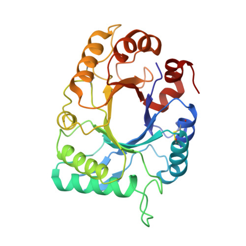
    A novel plant protein isolated from the underground bulbs of Scadoxus multiflorus, xylanase and alpha-amylase inhibitor protein (XAIP), inhibits two structurally and functionally unrelated enzymes: xylanase and alpha-amylase. The mature protein contains 272 amino acid residues which show sequence identities of 48% to the plant chitinase hevamine and 36% to xylanase inhibitor protein-I, a double-headed inhibitor of GH10 and GH11 xylanases. However, unlike hevamine, it is enzymatically inactive and, unlike xylanase inhibitor protein-I, it inhibits two functionally different classes of enzyme. The crystal structure of XAIP has been determined at 2.0 A resolution and refined to R(cryst) and R(free) factors of 15.2% and 18.6%, respectively. The polypeptide chain of XAIP adopts a modified triosephosphate isomerase barrel fold with eight beta-strands in the inner circle and nine alpha-helices forming the outer ring. The structure contains three cis peptide bonds: Gly33-Phe34, Tyr159-Pro160 and Trp253-Asp254. Although hevamine has a long accessible carbohydrate-binding channel, in XAIP this channel is almost completely filled with the side-chains of residues Phe13, Pro77, Lys78 and Trp253. Solution studies indicate that XAIP inhibits GH11 family xylanases and GH13 family alpha-amylases through two independent binding sites located on opposite surfaces of the protein. Comparison of the structure of XAIP with that of xylanase inhibitor protein-I, and docking studies, suggest that loops alpha3-beta4 and alpha4-beta5 may be involved in the binding of GH11 xylanase, and that helix alpha7 and loop beta6-alpha6 are suitable for the interaction with alpha-amylase.


  • Organizational Affiliation
    • Department of Biophysics, All India Institute of Medical Sciences, New Delhi, India.

Macromolecule Content 

  • Total Structure Weight: 30.31 kDa 
  • Atom Count: 2,255 
  • Modeled Residue Count: 272 
  • Deposited Residue Count: 272 
  • Unique protein chains: 1

Macromolecules

Find similar proteins by:|  3D Structure
Entity ID: 1
MoleculeChains  Sequence LengthOrganismDetailsImage
Haementhin272Scadoxus multiflorusMutation(s): 0 
EC: 3.2.1.14
UniProt
Find proteins for B2ZGS7 (Scadoxus multiflorus)
Explore B2ZGS7 
Go to UniProtKB:  B2ZGS7
Entity Groups
Sequence Clusters30% Identity50% Identity70% Identity90% Identity95% Identity100% Identity
UniProt GroupB2ZGS7
Sequence Annotations
Expand
Reference Sequence

Oligosaccharides

Help  
Entity ID: 2
MoleculeChains Length2D Diagram GlycosylationD Interactions
beta-D-glucopyranose-(1-4)-beta-D-glucopyranose
B
2N/A
Glycosylation Resources
GlyTouCan: G84824ZO
GlyCosmos: G84824ZO

Biologically Interesting Molecules (External Reference) 

1 Unique

Experimental Data & Validation

Experimental Data

  • Method: X-RAY DIFFRACTION
  • Resolution: 2.40 Å
  • R-Value Free:  0.212 (Depositor), 0.227 (DCC) 
  • R-Value Work:  0.196 (Depositor), 0.196 (DCC) 
  • R-Value Observed: 0.196 (Depositor) 
Space Group: P 1 21 1
Unit Cell:
Length ( Å )Angle ( ˚ )
a = 42.8α = 90
b = 65.6β = 102.1
c = 49.42γ = 90
Software Package:
Software NamePurpose
MAR345dtbdata collection
AMoREphasing
CNSrefinement
DENZOdata reduction
SCALEPACKdata scaling

Structure Validation

View Full Validation Report


Entry History 

Deposition Data

Revision History  (Full details and data files)

  • Version 1.0: 2010-05-05
    Type: Initial release
  • Version 1.1: 2011-07-13
    Changes: Version format compliance
  • Version 1.2: 2011-09-07
    Changes: Database references
  • Version 2.0: 2020-07-29
    Type: Remediation
    Reason: Carbohydrate remediation
    Changes: Atomic model, Data collection, Derived calculations, Non-polymer description, Structure summary
  • Version 2.1: 2023-11-01
    Changes: Data collection, Database references, Refinement description, Structure summary
  • Version 2.2: 2024-11-13
    Changes: Structure summary
  • Version 2.3: 2025-05-07
    Changes: Other