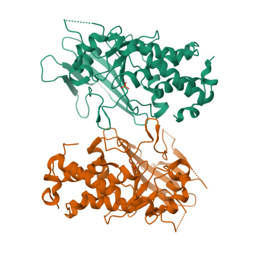
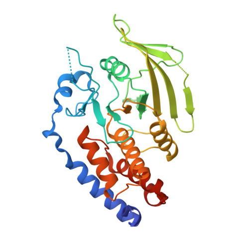
3M4U

Crystal Structure of Trypanosoma brucei Protein Tyrosine Phosphatase TbPTP1


Experimental Data Snapshot

  • Method: X-RAY DIFFRACTION
  • Resolution: 2.39 Å
  • R-Value Free: 0.256 
  • R-Value Work: 0.199 
  • R-Value Observed: 0.201 

wwPDB Validation   3D Report Full Report


This is version 1.3 of the entry. See complete history


Literature

The Trypanosoma brucei life cycle switch TbPTP1 is structurally conserved and dephosphorylates the nucleolar protein NOPP44/46.

Chou, S.Jensen, B.C.Parsons, M.Alber, T.Grundner, C.

(2010) J Biol Chem 285: 22075-22081

  • DOI: https://doi.org/10.1074/jbc.M110.108860
  • Primary Citation of Related Structures:  
    3M4U

  • PubMed Abstract: 

    Trypanosoma brucei adapts to changing environments as it cycles through arrested and proliferating stages in the human and tsetse fly hosts. Changes in protein tyrosine phosphorylation of several proteins, including NOPP44/46, accompany T. brucei development. Moreover, inactivation of T. brucei protein-tyrosine phosphatase 1 (TbPTP1) triggers differentiation of bloodstream stumpy forms into tsetse procyclic forms through unknown downstream effects. Here, we link these events by showing that NOPP44/46 is a major substrate of TbPTP1. TbPTP1 substrate-trapping mutants selectively enrich NOPP44/46 from procyclic stage cell lysates, and TbPTP1 efficiently and selectively dephosphorylates NOPP44/46 in vitro. To provide insights into the mechanism of NOPP44/46 recognition, we determined the crystal structure of TbPTP1. The TbPTP1 structure, the first of a kinetoplastid protein-tyrosine phosphatase (PTP), emphasizes the conservation of the PTP fold, extending to one of the most diverged eukaryotes. The structure reveals surfaces that may mediate substrate specificity and affords a template for the design of selective inhibitors to interfere with T. brucei transmission.


  • Organizational Affiliation

    Department of Molecular and Cell Biology and QB3 Institute, University of California, Berkeley, California 94720-3200, USA.


Macromolecules
Find similar proteins by:  (by identity cutoff)  |  3D Structure
Entity ID: 1
MoleculeChains Sequence LengthOrganismDetailsImage
Tyrosine specific protein phosphatase, putative
A, B
306Trypanosoma bruceiMutation(s): 0 
Gene Names: Tb10.70.0070
EC: 3.1.3.48
UniProt
Find proteins for Q38AT7 (Trypanosoma brucei brucei (strain 927/4 GUTat10.1))
Explore Q38AT7 
Go to UniProtKB:  Q38AT7
Entity Groups  
Sequence Clusters30% Identity50% Identity70% Identity90% Identity95% Identity100% Identity
UniProt GroupQ38AT7
Sequence Annotations
Expand
  • Reference Sequence
Experimental Data & Validation

Experimental Data

  • Method: X-RAY DIFFRACTION
  • Resolution: 2.39 Å
  • R-Value Free: 0.256 
  • R-Value Work: 0.199 
  • R-Value Observed: 0.201 
  • Space Group: P 21 21 21
Unit Cell:
Length ( Å )Angle ( ˚ )
a = 76.632α = 90
b = 77.377β = 90
c = 117.195γ = 90
Software Package:
Software NamePurpose
DENZOdata reduction
SCALEPACKdata scaling
MOLREPphasing
PHENIXrefinement
PDB_EXTRACTdata extraction

Structure Validation

View Full Validation Report


Entry History 

Deposition Data

Revision History  (Full details and data files)

  • Version 1.0: 2010-05-05
    Type: Initial release
  • Version 1.1: 2011-07-13
    Changes: Version format compliance
  • Version 1.2: 2017-11-08
    Changes: Refinement description
  • Version 1.3: 2024-02-21
    Changes: Data collection, Database references, Derived calculations