3M4E | pdb_00003m4e

Crystal structure of the M113N mutant of alpha-hemolysin bound to beta-cyclodextrin


Experimental Data Snapshot

  • Method: X-RAY DIFFRACTION
  • Resolution: 2.30 Å
  • R-Value Free: 
    0.269 (Depositor), 0.244 (DCC) 
  • R-Value Work: 
    0.229 (Depositor), 0.232 (DCC) 

wwPDB Validation 3D Report Full Report

Validation slider image for 3M4E

This is version 2.2 of the entry. See complete history

Literature

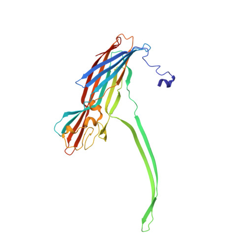
Molecular bases of cyclodextrin adapter interactions with engineered protein nanopores.

Banerjee, A.Mikhailova, E.Cheley, S.Gu, L.Q.Montoya, M.Nagaoka, Y.Gouaux, E.Bayley, H.

(2010) Proc Natl Acad Sci U S A 107: 8165-8170

  • DOI: https://doi.org/10.1073/pnas.0914229107
  • Primary Citation Related Structures: 
    3M2L, 3M3R, 3M4D, 3M4E

  • PubMed Abstract: 

    Engineered protein pores have several potential applications in biotechnology: as sensor elements in stochastic detection and ultrarapid DNA sequencing, as nanoreactors to observe single-molecule chemistry, and in the construction of nano- and micro-devices. One important class of pores contains molecular adapters, which provide internal binding sites for small molecules. Mutants of the alpha-hemolysin (alphaHL) pore that bind the adapter beta-cyclodextrin (betaCD) approximately 10(4) times more tightly than the wild type have been obtained. We now use single-channel electrical recording, protein engineering including unnatural amino acid mutagenesis, and high-resolution x-ray crystallography to provide definitive structural information on these engineered protein nanopores in unparalleled detail.


  • Organizational Affiliation
    • Department of Chemistry, University of Oxford, Oxford, OX1 3TA, United Kingdom.

Macromolecule Content 

  • Total Structure Weight: 234.06 kDa 
  • Atom Count: 16,774 
  • Modeled Residue Count: 2,051 
  • Deposited Residue Count: 2,051 
  • Unique protein chains: 1

Macromolecules

Find similar proteins by:|  3D Structure
Entity ID: 1
MoleculeChains  Sequence LengthOrganismDetailsImage
Alpha-hemolysin
A, B, C, D, E
A, B, C, D, E, F, G
293Staphylococcus aureusMutation(s): 1 
Gene Names: hlahly
Membrane Entity: Yes 
UniProt
Find proteins for P09616 (Staphylococcus aureus)
Explore P09616 
Go to UniProtKB:  P09616
Entity Groups
Sequence Clusters30% Identity50% Identity70% Identity90% Identity95% Identity100% Identity
UniProt GroupP09616
Sequence Annotations
Expand
Reference Sequence

Oligosaccharides

Help  
Entity ID: 2
MoleculeChains Length2D Diagram GlycosylationD Interactions
Cycloheptakis-(1-4)-(alpha-D-glucopyranose)
H
7N/A
Glycosylation Resources
GlyTouCan: G01435GL
GlyCosmos: G01435GL

Biologically Interesting Molecules (External Reference) 

1 Unique

Experimental Data & Validation

Experimental Data

  • Method: X-RAY DIFFRACTION
  • Resolution: 2.30 Å
  • R-Value Free:  0.269 (Depositor), 0.244 (DCC) 
  • R-Value Work:  0.229 (Depositor), 0.232 (DCC) 
Space Group: C 1 2 1
Unit Cell:
Length ( Å )Angle ( ˚ )
a = 150.35α = 90
b = 134.27β = 90.97
c = 132.06γ = 90
Software Package:
Software NamePurpose
HKL-2000data collection
CNSrefinement
DENZOdata reduction
SCALEPACKdata scaling
CNSphasing

Structure Validation

View Full Validation Report


Entry History 

Deposition Data

Revision History  (Full details and data files)

  • Version 1.0: 2010-05-05
    Type: Initial release
  • Version 1.1: 2011-07-13
    Changes: Version format compliance
  • Version 2.0: 2020-07-29
    Type: Remediation
    Reason: Carbohydrate remediation
    Changes: Advisory, Atomic model, Data collection, Derived calculations, Non-polymer description, Structure summary
  • Version 2.1: 2021-10-06
    Changes: Advisory, Database references, Structure summary
  • Version 2.2: 2024-02-21
    Changes: Data collection