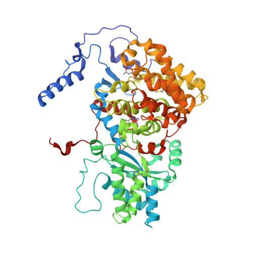
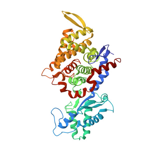
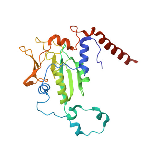
3M1V

Structural Insight into Methyl-Coenzyme M Reductase Chemistry using Coenzyme B Analogues


Experimental Data Snapshot

  • Method: X-RAY DIFFRACTION
  • Resolution: 1.45 Å
  • R-Value Free: 0.162 
  • R-Value Work: 0.135 
  • R-Value Observed: 0.136 

wwPDB Validation   3D Report Full Report


Ligand Structure Quality Assessment 


This is version 1.4 of the entry. See complete history


Literature

Structural insight into methyl-coenzyme M reductase chemistry using coenzyme B analogues.

Cedervall, P.E.Dey, M.Pearson, A.R.Ragsdale, S.W.Wilmot, C.M.

(2010) Biochemistry 49: 7683-7693

  • DOI: https://doi.org/10.1021/bi100458d
  • Primary Citation of Related Structures:  
    3M1V, 3M2R, 3M2U, 3M2V, 3M30, 3M32

  • PubMed Abstract: 

    Methyl-coenzyme M reductase (MCR) catalyzes the final and rate-limiting step in methane biogenesis: the reduction of methyl-coenzyme M (methyl-SCoM) by coenzyme B (CoBSH) to methane and a heterodisulfide (CoBS-SCoM). Crystallographic studies show that the active site is deeply buried within the enzyme and contains a highly reduced nickel-tetrapyrrole, coenzyme F(430). Methyl-SCoM must enter the active site prior to CoBSH, as species derived from methyl-SCoM are always observed bound to the F(430) nickel in the deepest part of the 30 A long substrate channel that leads from the protein surface to the active site. The seven-carbon mercaptoalkanoyl chain of CoBSH binds within a 16 A predominantly hydrophobic part of the channel close to F(430), with the CoBSH thiolate lying closest to the nickel at a distance of 8.8 A. It has previously been suggested that binding of CoBSH initiates catalysis by inducing a conformational change that moves methyl-SCoM closer to the nickel promoting cleavage of the C-S bond of methyl-SCoM. In order to better understand the structural role of CoBSH early in the MCR mechanism, we have determined crystal structures of MCR in complex with four different CoBSH analogues: pentanoyl, hexanoyl, octanoyl, and nonanoyl derivatives of CoBSH (CoB(5)SH, CoB(6)SH, CoB(8)SH, and CoB(9)SH, respectively). The data presented here reveal that the shorter CoB(5)SH mercaptoalkanoyl chain overlays with that of CoBSH but terminates two units short of the CoBSH thiolate position. In contrast, the mercaptoalkanoyl chain of CoB(6)SH adopts a different conformation, such that its thiolate is coincident with the position of the CoBSH thiolate. This is consistent with the observation that CoB(6)SH is a slow substrate. A labile water in the substrate channel was found to be a sensitive indicator for the presence of CoBSH and HSCoM. The longer CoB(8)SH and CoB(9)SH analogues can be accommodated in the active site through exclusion of this water. These analogues react with Ni(III)-methyl, a proposed MCR catalytic intermediate of methanogenesis. The CoB(8)SH thiolate is 2.6 A closer to the nickel than that of CoBSH, but the additional carbon of CoB(9)SH only decreases the nickel thiolate distance a further 0.3 A. Although the analogues do not induce any structural changes in the substrate channel, the thiolates appear to preferentially bind at two distinct positions in the channel, one being the previously observed CoBSH thiolate position and the other being at a hydrophobic annulus of residues that lines the channel proximal to the nickel.


  • Organizational Affiliation

    Department of Biochemistry, Molecular Biology, and Biophysics, University of Minnesota, Minneapolis, Minnesota 55455, USA.


Macromolecules
Find similar proteins by:  (by identity cutoff)  |  3D Structure
Entity ID: 1
MoleculeChains Sequence LengthOrganismDetailsImage
Methyl-coenzyme M reductase I subunit alpha
A, D
549Methanothermobacter marburgensis str. MarburgMutation(s): 0 
EC: 2.8.4.1
UniProt
Find proteins for P11558 (Methanothermobacter marburgensis (strain ATCC BAA-927 / DSM 2133 / JCM 14651 / NBRC 100331 / OCM 82 / Marburg))
Explore P11558 
Go to UniProtKB:  P11558
Entity Groups  
Sequence Clusters30% Identity50% Identity70% Identity90% Identity95% Identity100% Identity
UniProt GroupP11558
Sequence Annotations
Expand
  • Reference Sequence
Find similar proteins by:  (by identity cutoff)  |  3D Structure
Entity ID: 2
MoleculeChains Sequence LengthOrganismDetailsImage
Methyl-coenzyme M reductase I subunit beta
B, E
442Methanothermobacter marburgensis str. MarburgMutation(s): 0 
EC: 2.8.4.1
UniProt
Find proteins for P11560 (Methanothermobacter marburgensis (strain ATCC BAA-927 / DSM 2133 / JCM 14651 / NBRC 100331 / OCM 82 / Marburg))
Explore P11560 
Go to UniProtKB:  P11560
Entity Groups  
Sequence Clusters30% Identity50% Identity70% Identity90% Identity95% Identity100% Identity
UniProt GroupP11560
Sequence Annotations
Expand
  • Reference Sequence
Find similar proteins by:  (by identity cutoff)  |  3D Structure
Entity ID: 3
MoleculeChains Sequence LengthOrganismDetailsImage
Methyl-coenzyme M reductase I subunit gamma
C, F
248Methanothermobacter marburgensis str. MarburgMutation(s): 0 
EC: 2.8.4.1
UniProt
Find proteins for P11562 (Methanothermobacter marburgensis (strain ATCC BAA-927 / DSM 2133 / JCM 14651 / NBRC 100331 / OCM 82 / Marburg))
Explore P11562 
Go to UniProtKB:  P11562
Entity Groups  
Sequence Clusters30% Identity50% Identity70% Identity90% Identity95% Identity100% Identity
UniProt GroupP11562
Sequence Annotations
Expand
  • Reference Sequence
Small Molecules
Ligands 8 Unique
IDChains Name / Formula / InChI Key2D Diagram3D Interactions
F43
Query on F43

Download Ideal Coordinates CCD File 
I [auth A],
X [auth D]
FACTOR 430
C42 H51 N6 Ni O13
XLFIRMYGVLUNOY-SXMZNAGASA-M
TP7
Query on TP7

Download Ideal Coordinates CCD File 
J [auth A],
Y [auth D]
Coenzyme B
C11 H22 N O7 P S
JBJSVEVEEGOEBZ-SCZZXKLOSA-N
COM
Query on COM

Download Ideal Coordinates CCD File 
K [auth A],
Z [auth D]
1-THIOETHANESULFONIC ACID
C2 H6 O3 S2
ZNEWHQLOPFWXOF-UHFFFAOYSA-N
PEG
Query on PEG

Download Ideal Coordinates CCD File 
N [auth A],
U [auth C]
DI(HYDROXYETHYL)ETHER
C4 H10 O3
MTHSVFCYNBDYFN-UHFFFAOYSA-N
ZN
Query on ZN

Download Ideal Coordinates CCD File 
O [auth A]ZINC ION
Zn
PTFCDOFLOPIGGS-UHFFFAOYSA-N
EDO
Query on EDO

Download Ideal Coordinates CCD File 
AA [auth D],
DA [auth F],
EA [auth F]
1,2-ETHANEDIOL
C2 H6 O2
LYCAIKOWRPUZTN-UHFFFAOYSA-N
ACT
Query on ACT

Download Ideal Coordinates CCD File 
L [auth A],
M [auth A],
R [auth B],
T [auth C]
ACETATE ION
C2 H3 O2
QTBSBXVTEAMEQO-UHFFFAOYSA-M
MG
Query on MG

Download Ideal Coordinates CCD File 
BA [auth E]
CA [auth F]
G [auth A]
H [auth A]
P [auth B]
BA [auth E],
CA [auth F],
G [auth A],
H [auth A],
P [auth B],
Q [auth B],
S [auth C],
V [auth D],
W [auth D]
MAGNESIUM ION
Mg
JLVVSXFLKOJNIY-UHFFFAOYSA-N
Modified Residues  5 Unique
IDChains TypeFormula2D DiagramParent
AGM
Query on AGM
A, D
L-PEPTIDE LINKINGC7 H17 N4 O2ARG
GL3
Query on GL3
A, D
L-PEPTIDE LINKINGC2 H5 N O SGLY
MGN
Query on MGN
A, D
L-PEPTIDE LINKINGC6 H12 N2 O3GLN
MHS
Query on MHS
A, D
L-PEPTIDE LINKINGC7 H11 N3 O2HIS
SMC
Query on SMC
A, D
L-PEPTIDE LINKINGC4 H9 N O2 SCYS
Experimental Data & Validation

Experimental Data

  • Method: X-RAY DIFFRACTION
  • Resolution: 1.45 Å
  • R-Value Free: 0.162 
  • R-Value Work: 0.135 
  • R-Value Observed: 0.136 
  • Space Group: P 1 21 1
Unit Cell:
Length ( Å )Angle ( ˚ )
a = 82.016α = 90
b = 118.26β = 91.84
c = 122.387γ = 90
Software Package:
Software NamePurpose
DENZOdata reduction
SCALEPACKdata scaling
REFMACrefinement
PDB_EXTRACTdata extraction
BioCARS-developeddata collection
REFMACphasing

Structure Validation

View Full Validation Report


Ligand Structure Quality Assessment 


Entry History 

Deposition Data

Revision History  (Full details and data files)

  • Version 1.0: 2010-09-15
    Type: Initial release
  • Version 1.1: 2011-07-13
    Changes: Version format compliance
  • Version 1.2: 2015-05-20
    Changes: Non-polymer description
  • Version 1.3: 2017-03-29
    Changes: Non-polymer description
  • Version 1.4: 2017-11-08
    Changes: Refinement description