3M1B | pdb_00003m1b

Crystal structure of human FcRn with a dimeric peptide inhibitor


Experimental Data Snapshot

  • Method: X-RAY DIFFRACTION
  • Resolution: 3.10 Å
  • R-Value Free: 
    0.397 (Depositor), 0.395 (DCC) 
  • R-Value Work: 
    0.314 (Depositor), 0.306 (DCC) 
  • R-Value Observed: 
    0.318 (Depositor) 

Starting Model: experimental
View more details

wwPDB Validation 3D Report Full Report

Validation slider image for 3M1B

This is version 1.6 of the entry. See complete history

Literature

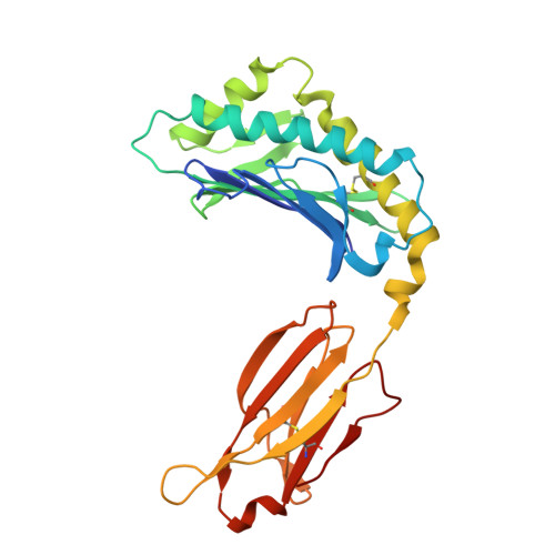
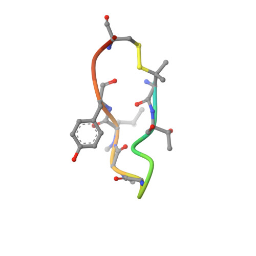
X-ray crystal structures of monomeric and dimeric peptide inhibitors in complex with the human neonatal Fc receptor, FcRn.

Mezo, A.R.Sridhar, V.Badger, J.Sakorafas, P.Nienaber, V.

(2010) J Biological Chem 285: 27694-27701

  • DOI: https://doi.org/10.1074/jbc.M110.120667
  • Primary Citation Related Structures: 
    3M17, 3M1B

  • PubMed Abstract: 

    The neonatal Fc receptor, FcRn, is responsible for the long half-life of IgG molecules in vivo and is a potential therapeutic target for the treatment of autoimmune diseases. A family of peptides comprising the consensus motif GHFGGXY, where X is preferably a hydrophobic amino acid, was shown previously to inhibit the human IgG:human FcRn protein-protein interaction (Mezo, A. R., McDonnell, K. A., Tan Hehir, C. A., Low, S. C., Palombella, V. J., Stattel, J. M., Kamphaus, G. D., Fraley, C., Zhang, Y., Dumont, J. A., and Bitonti, A. J. (2008) Proc. Natl. Acad. Sci. U.S.A., 105, 2337-2342). Herein, the x-ray crystal structure of a representative monomeric peptide in complex with human FcRn was solved to 2.6 A resolution. The structure shows that the peptide binds to human FcRn at the same general binding site as does the Fc domain of IgG. The data correlate well with structure-activity relationship data relating to how the peptide family binds to human FcRn. In addition, the x-ray crystal structure of a representative dimeric peptide in complex with human FcRn shows how the bivalent ligand can bridge two FcRn molecules, which may be relevant to the mechanism by which the dimeric peptides inhibit FcRn and increase IgG catabolism in vivo. Modeling of the peptide:FcRn structure as compared with available structural data on Fc and FcRn suggest that the His-6 and Phe-7 (peptide) partially mimic the interaction of His-310 and Ile-253 (Fc) in binding to FcRn, but using a different backbone topology.


  • Organizational Affiliation
    • Biogen Idec, Syntonix Subsidiary, Waltham, Massachusetts 02451, USA. adam.mezo@biogenidec.com

Macromolecule Content 

  • Total Structure Weight: 169.21 kDa 
  • Atom Count: 11,241 
  • Modeled Residue Count: 1,471 
  • Deposited Residue Count: 1,496 
  • Unique protein chains: 3

Macromolecules

Find similar proteins by:|  3D Structure
Entity ID: 1
MoleculeChains  Sequence LengthOrganismDetailsImage
IgG receptor FcRn large subunit p51
A, C, E, G
267Homo sapiensMutation(s): 0 
Gene Names: FCGRTFCRN
UniProt & NIH Common Fund Data Resources
Find proteins for P55899 (Homo sapiens)
Explore P55899 
Go to UniProtKB:  P55899
PHAROS:  P55899
GTEx:  ENSG00000104870 
Entity Groups
Sequence Clusters30% Identity50% Identity70% Identity90% Identity95% Identity100% Identity
UniProt GroupP55899
Sequence Annotations
Expand
Reference Sequence
Find similar proteins by:|  3D Structure
Entity ID: 2
MoleculeChains  Sequence LengthOrganismDetailsImage
Beta-2-microglobulin
B, D, F, H
99Homo sapiensMutation(s): 0 
Gene Names: B2MCDABP0092HDCMA22P
UniProt & NIH Common Fund Data Resources
Find proteins for P61769 (Homo sapiens)
Explore P61769 
Go to UniProtKB:  P61769
PHAROS:  P61769
GTEx:  ENSG00000166710 
Entity Groups
Sequence Clusters30% Identity50% Identity70% Identity90% Identity95% Identity100% Identity
UniProt GroupP61769
Sequence Annotations
Expand
Reference Sequence
Find similar proteins by:  Sequence   |   3D Structure  
Entity ID: 3
MoleculeChains  Sequence LengthOrganismDetailsImage
DIMERIC PEPTIDE INHIBITOR
I, J
16N/AMutation(s): 0 

Small Molecules

Modified Residues  3 Unique
IDChains TypeFormula2D DiagramParent
LE1
Query on LE1
I, J
L-PEPTIDE LINKINGC5 H11 N O2 SVAL
MLE
Query on MLE
I, J
L-PEPTIDE LINKINGC7 H15 N O2LEU
SAR
Query on SAR
I, J
PEPTIDE LINKINGC3 H7 N O2GLY

Biologically Interesting Molecules (External Reference) 

1 Unique

Experimental Data & Validation

Experimental Data

  • Method: X-RAY DIFFRACTION
  • Resolution: 3.10 Å
  • R-Value Free:  0.397 (Depositor), 0.395 (DCC) 
  • R-Value Work:  0.314 (Depositor), 0.306 (DCC) 
  • R-Value Observed: 0.318 (Depositor) 
Space Group: P 1 21 1
Unit Cell:
Length ( Å )Angle ( ˚ )
a = 68.05α = 90
b = 158.431β = 90.11
c = 82.539γ = 90
Software Package:
Software NamePurpose
PHASERphasing
REFMACrefinement
HKL-2000data reduction
HKL-2000data scaling

Structure Validation

View Full Validation Report


Entry History 

Deposition Data

Revision History  (Full details and data files)

  • Version 1.0: 2010-06-16
    Type: Initial release
  • Version 1.1: 2011-07-13
    Changes: Version format compliance
  • Version 1.2: 2011-08-24
    Changes: Derived calculations
  • Version 1.3: 2012-12-12
    Changes: Other
  • Version 1.4: 2022-05-04
    Changes: Database references, Derived calculations, Structure summary
  • Version 1.5: 2023-09-06
    Changes: Data collection, Refinement description
  • Version 1.6: 2023-11-22
    Changes: Data collection