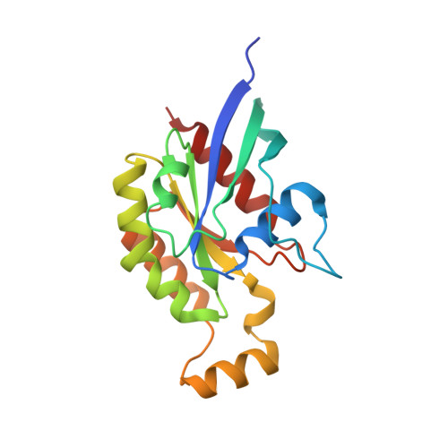
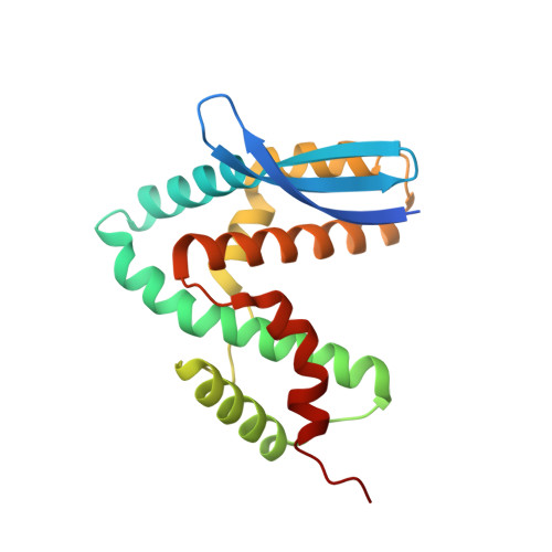
3LWN

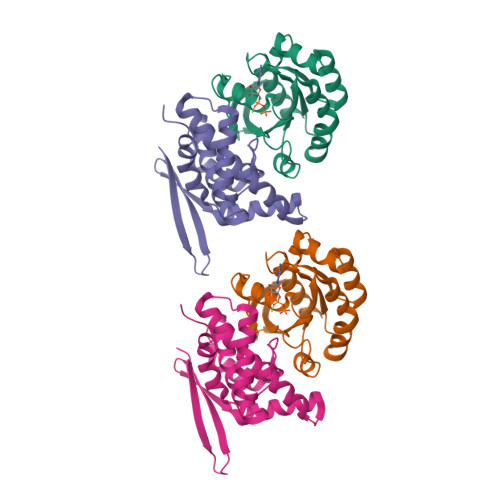
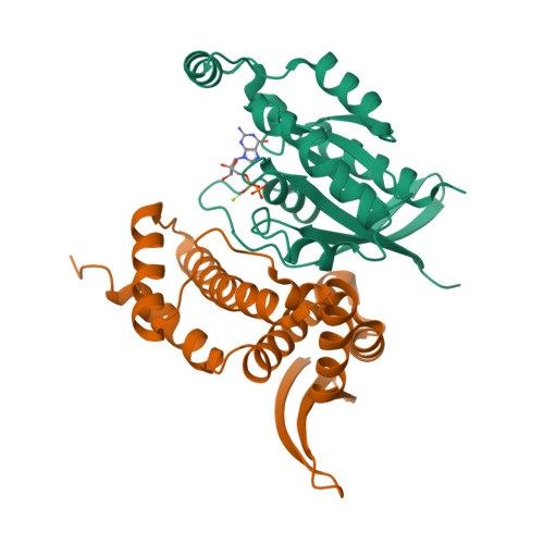
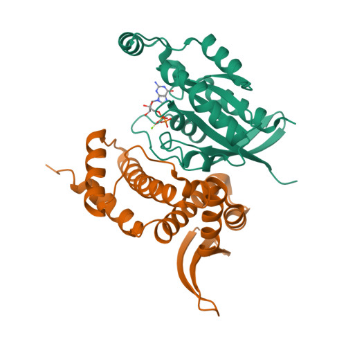
Shigella IpgB2 in complex with human RhoA, GDP and Mg2+ (complex B)


Experimental Data Snapshot

  • Method: X-RAY DIFFRACTION
  • Resolution: 2.28 Å
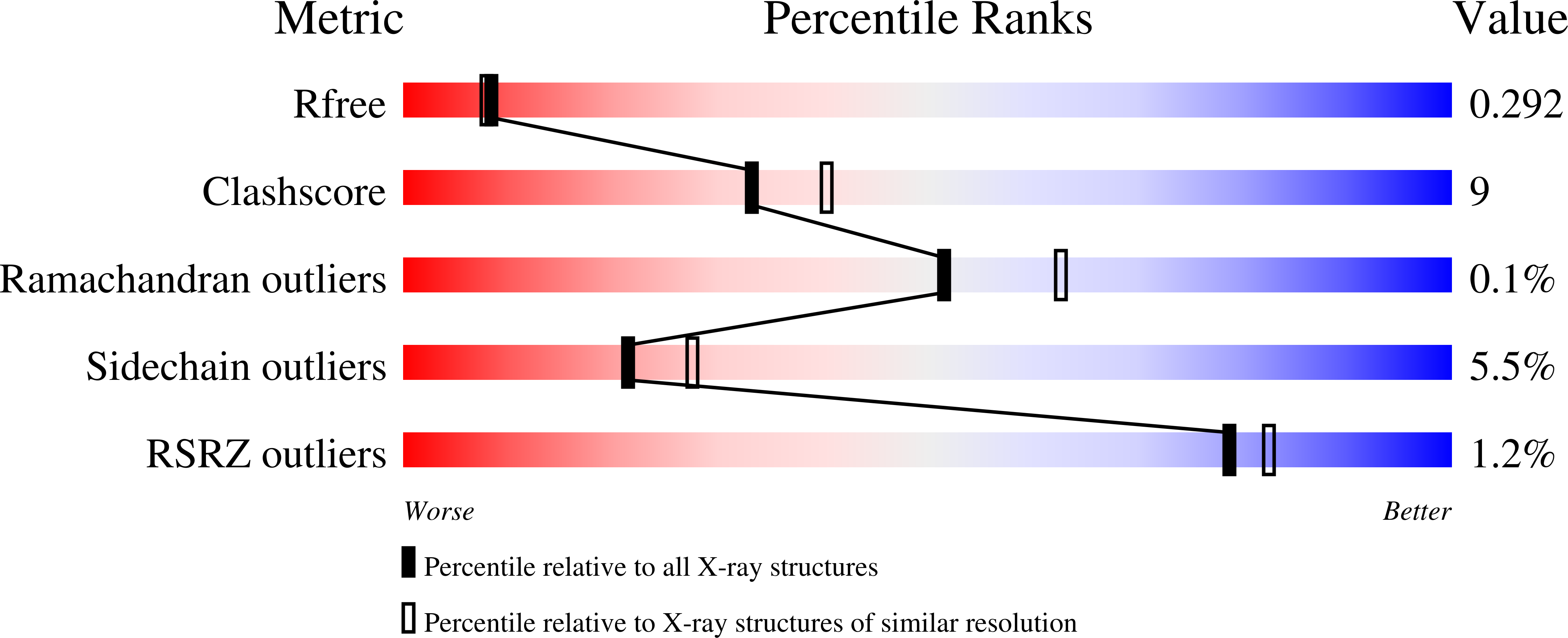
  • R-Value Free: 0.291 
  • R-Value Work: 0.204 
  • R-Value Observed: 0.209 

Starting Model: experimental
View more details

wwPDB Validation   3D Report Full Report


Ligand Structure Quality Assessment 


This is version 1.2 of the entry. See complete history


Literature

Structure of Shigella IPGB2 in complex with human RhoA: Implications for the mechanism of bacterial GEF-mimicry

Klink, B.U.Barden, S.Heidler, T.V.Borchers, C.Ladwein, M.Stradal, T.E.B.Rottner, K.Heinz, D.W.

(2010) J Biol Chem 285: 17197-17208

  • DOI: https://doi.org/10.1074/jbc.M110.107953
  • Primary Citation of Related Structures:  
    3LW8, 3LWN, 3LXR, 3LYQ

  • PubMed Abstract: 

    A common theme in bacterial pathogenesis is the manipulation of eukaryotic cells by targeting the cytoskeleton. This is in most cases achieved either by modifying actin, or indirectly via activation of key regulators controlling actin dynamics such as Rho-GTPases. A novel group of bacterial virulence factors termed the WXXXE family has emerged as guanine nucleotide exchange factors (GEFs) for these GTPases. The precise mechanism of nucleotide exchange, however, has remained unclear. Here we report the structure of the WXXXE-protein IpgB2 from Shigella flexneri and its complex with human RhoA. We unambiguously identify IpgB2 as a bacterial RhoA-GEF and dissect the molecular mechanism of GDP release, an essential prerequisite for GTP binding. Our observations uncover that IpgB2 induces conformational changes on RhoA mimicking DbI- but not DOCK family GEFs. We also show that dissociation of the GDP.Mg(2+) complex is preceded by the displacement of the metal ion to the alpha-phosphate of the nucleotide, diminishing its affinity to the GTPase. These data refine our understanding of the mode of action not only of WXXXE GEFs but also of mammalian GEFs of the DH/PH family.


  • Organizational Affiliation

    Division of Structural Biology, Helmholtz Zentrum für Infektionsforschung, D-38124 Braunschweig, Germany.


Macromolecules
Find similar proteins by:  (by identity cutoff)  |  3D Structure
Entity ID: 1
MoleculeChains Sequence LengthOrganismDetailsImage
Transforming protein RhoA
A, B
185Homo sapiensMutation(s): 0 
Gene Names: RHOA
EC: 3.6.5.2
UniProt & NIH Common Fund Data Resources
Find proteins for P61586 (Homo sapiens)
Explore P61586 
Go to UniProtKB:  P61586
PHAROS:  P61586
GTEx:  ENSG00000067560 
Entity Groups  
Sequence Clusters30% Identity50% Identity70% Identity90% Identity95% Identity100% Identity
UniProt GroupP61586
Sequence Annotations
Expand
  • Reference Sequence
Find similar proteins by:  (by identity cutoff)  |  3D Structure
Entity ID: 2
MoleculeChains Sequence LengthOrganismDetailsImage
IpgB2C [auth F],
D [auth G]
192Shigella flexneriMutation(s): 0 
Gene Names: ipgB2
UniProt
Find proteins for Q9AJW7 (Shigella flexneri)
Explore Q9AJW7 
Go to UniProtKB:  Q9AJW7
Entity Groups  
Sequence Clusters30% Identity50% Identity70% Identity90% Identity95% Identity100% Identity
UniProt GroupQ9AJW7
Sequence Annotations
Expand
  • Reference Sequence
Experimental Data & Validation

Experimental Data

  • Method: X-RAY DIFFRACTION
  • Resolution: 2.28 Å
  • R-Value Free: 0.291 
  • R-Value Work: 0.204 
  • R-Value Observed: 0.209 
  • Space Group: P 21 21 21
Unit Cell:
Length ( Å )Angle ( ˚ )
a = 75.53α = 90
b = 95.7β = 90
c = 102.96γ = 90
Software Package:
Software NamePurpose
XSCALEdata scaling
MOLREPphasing
REFMACrefinement
PDB_EXTRACTdata extraction

Structure Validation

View Full Validation Report


Ligand Structure Quality Assessment 


Entry History 

Deposition Data

Revision History  (Full details and data files)

  • Version 1.0: 2010-03-31
    Type: Initial release
  • Version 1.1: 2011-07-13
    Changes: Advisory, Refinement description, Version format compliance
  • Version 1.2: 2023-11-01
    Changes: Data collection, Database references, Derived calculations, Refinement description