3LU6 | pdb_00003lu6

Human serum albumin in complex with compound 1


Experimental Data Snapshot

  • Method: X-RAY DIFFRACTION
  • Resolution: 2.70 Å
  • R-Value Free: 
    0.275 (Depositor), 0.277 (DCC) 
  • R-Value Work: 
    0.236 (Depositor), 0.235 (DCC) 
  • R-Value Observed: 
    0.238 (Depositor) 

wwPDB Validation 3D Report Full Report

Validation slider image for 3LU6

Ligand Structure Quality Assessment 


This is version 1.2 of the entry. See complete history

Literature

A combined spectroscopic and crystallographic approach to probing drug-human serum albumin interactions

Buttar, D.Colclough, N.Gerhardt, S.Macfaul, P.A.Phillips, S.D.Plowright, A.Whittamore, P.Tam, K.Maskos, K.Steinbacher, S.Steuber, H.

(2010) Bioorg Med Chem 18: 7486-7496

  • DOI: https://doi.org/10.1016/j.bmc.2010.08.052
  • Primary Citation Related Structures: 
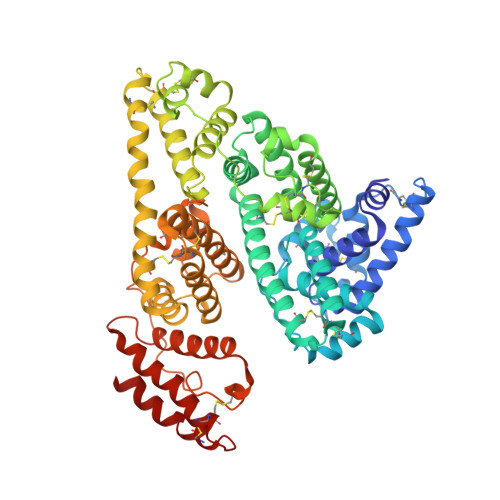
    3LU6, 3LU7, 3LU8

  • PubMed Abstract: 

    The displacement of probes that bind selectively to subdomains IIA or IIIA on human serum albumin (HSA) by competing compounds has been followed using fluorescence spectroscopy, and has therefore been used to assign a primary binding site for these compounds in the presence and absence of fatty acids. The crystal structures have also been solved for three compounds: a matched pair of carboxylic acids whose binding strength to HSA unexpectedly decreased as the lipophilicity increased; and a highly bound sulphonamide that appeared not to displace the probes in the displacement assay. The crystallography results support the findings from the fluorescence displacement assay. The results indicate that drug binding to subdomain IB might also be important location for certain compounds.


  • Organizational Affiliation
    • AstraZeneca, Alderley Park, Macclesfield, Cheshire SK104TG, England, United Kingdom.

Macromolecule Content 

  • Total Structure Weight: 134.55 kDa 
  • Atom Count: 9,322 
  • Modeled Residue Count: 1,156 
  • Deposited Residue Count: 1,170 
  • Unique protein chains: 1

Macromolecules

Find similar proteins by:|  3D Structure
Entity ID: 1
MoleculeChains  Sequence LengthOrganismDetailsImage
Serum albumin
A, B
585Homo sapiensMutation(s): 0 
UniProt & NIH Common Fund Data Resources
Find proteins for P02768 (Homo sapiens)
Explore P02768 
Go to UniProtKB:  P02768
PHAROS:  P02768
GTEx:  ENSG00000163631 
Entity Groups
Sequence Clusters30% Identity50% Identity70% Identity90% Identity95% Identity100% Identity
UniProt GroupP02768
Sequence Annotations
Expand
Reference Sequence

Experimental Data & Validation

Experimental Data

  • Method: X-RAY DIFFRACTION
  • Resolution: 2.70 Å
  • R-Value Free:  0.275 (Depositor), 0.277 (DCC) 
  • R-Value Work:  0.236 (Depositor), 0.235 (DCC) 
  • R-Value Observed: 0.238 (Depositor) 
Space Group: P 1
Unit Cell:
Length ( Å )Angle ( ˚ )
a = 58.561α = 74.51
b = 59.868β = 86.57
c = 95.573γ = 74.47
Software Package:
Software NamePurpose
MOSFLMdata reduction
MOLREPphasing
REFMACrefinement
SCALAdata scaling

Structure Validation

View Full Validation Report


Ligand Structure Quality Assessment 


Entry History 

Revision History  (Full details and data files)

  • Version 1.0: 2010-10-27
    Type: Initial release
  • Version 1.1: 2011-07-13
    Changes: Version format compliance
  • Version 1.2: 2024-11-13
    Changes: Data collection, Database references, Derived calculations, Structure summary