


Crystal structure of glutathione s-transferase from Rhodobacter sphaeroides
Eswaramoorthy, S., Burley, S.K., Swaminathan, S.To be published.
Experimental Data Snapshot
Entity ID: 1 | |||||
|---|---|---|---|---|---|
| Molecule | Chains | Sequence Length | Organism | Details | Image |
| Glutathione S-transferase | 225 | Cereibacter sphaeroides 2.4.1 | Mutation(s): 0 Gene Names: RHOS4_23800, RSP_0769 EC: 2.5.1.18 | 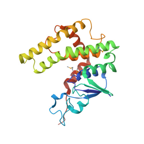 | |
UniProt | |||||
Entity Groups | |||||
| Sequence Clusters | 30% Identity50% Identity70% Identity90% Identity95% Identity100% Identity | ||||
| UniProt Group | Q3IZT6 | ||||
Sequence AnnotationsExpand | |||||
Reference Sequence | |||||
| Ligands 3 Unique | |||||
|---|---|---|---|---|---|
| ID | Chains | Name / Formula / InChI Key | 2D Diagram | 3D Interactions | |
| GSH Download:Ideal Coordinates CCD File | E [auth A], G [auth B], J [auth C], L [auth D] | Glutathione C10 H17 N3 O6 S RWSXRVCMGQZWBV-WDSKDSINSA-N |  | ||
| TRS Download:Ideal Coordinates CCD File | F [auth A], H [auth B], K [auth C], M [auth D] | 2-AMINO-2-HYDROXYMETHYL-PROPANE-1,3-DIOL C4 H12 N O3 LENZDBCJOHFCAS-UHFFFAOYSA-O |  | ||
| GOL Download:Ideal Coordinates CCD File | I [auth B], N [auth D] | GLYCEROL C3 H8 O3 PEDCQBHIVMGVHV-UHFFFAOYSA-N |  | ||
| Modified Residues 1 Unique | |||||
|---|---|---|---|---|---|
| ID | Chains | Type | Formula | 2D Diagram | Parent |
| MSE Query on MSE | A, B, C, D | L-PEPTIDE LINKING | C5 H11 N O2 Se |  | MET |
| Entity ID: 2 | |||||
|---|---|---|---|---|---|
| ID | Chains | Name | Type/Class | 2D Diagram | 3D Interactions |
| PRD_002593 (GSH) Query on PRD_002593 | E [auth A], G [auth B], J [auth C], L [auth D] | Glutathione | Peptide-like / Oxidation-reduction | | |
| Length ( Å ) | Angle ( ˚ ) |
|---|---|
| a = 167.92 | α = 90 |
| b = 50.398 | β = 125.51 |
| c = 133.228 | γ = 90 |
| Software Name | Purpose |
|---|---|
| CBASS | data collection |
| SHELXS | phasing |
| SHARP | phasing |
| CNS | refinement |
| HKL-2000 | data reduction |
| HKL-2000 | data scaling |


