3LS2 | pdb_00003ls2

Crystal structure of an S-formylglutathione hydrolase from Pseudoalteromonas haloplanktis TAC125


Experimental Data Snapshot

  • Method: X-RAY DIFFRACTION
  • Resolution: 2.20 Å
  • R-Value Free: 
    0.205 (Depositor), 0.206 (DCC) 
  • R-Value Work: 
    0.161 (Depositor), 0.161 (DCC) 

Starting Model: experimental
View more details

wwPDB Validation 3D Report Full Report

Validation slider image for 3LS2

This is version 1.2 of the entry. See complete history

Literature

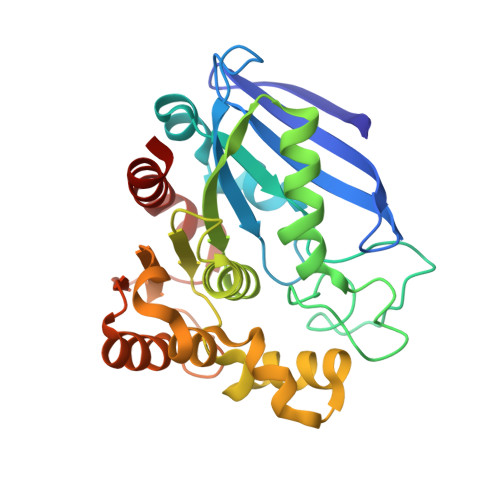
Crystal structure of an S-formylglutathione hydrolase from Pseudoalteromonas haloplanktis TAC125.

Alterio, V.Aurilia, V.Romanelli, A.Parracino, A.Saviano, M.D'Auria, S.De Simone, G.

(2010) Biopolymers 93: 669-677

  • DOI: https://doi.org/10.1002/bip.21420
  • Primary Citation Related Structures: 
    3LS2

  • PubMed Abstract: 

    S-formylglutathione hydrolases (FGHs) constitute a family of ubiquitous enzymes which play a key role in formaldehyde detoxification both in prokaryotes and eukaryotes, catalyzing the hydrolysis of S-formylglutathione to formic acid and glutathione. While a large number of functional studies have been reported on these enzymes, few structural studies have so far been carried out. In this article we report on the functional and structural characterization of PhEst, a FGH isolated from the psychrophilic bacterium Pseudoalteromonas haloplanktis. According to our functional studies, this enzyme is able to efficiently hydrolyze several thioester substrates with very small acyl moieties. By contrast, the enzyme shows no activity toward substrates with bulky acyl groups. These data are in line with structural studies which highlight for this enzyme a very narrow acyl-binding pocket in a typical alpha/beta-hydrolase fold. PhEst represents the first cold-adapted FGH structurally characterized to date; comparison with its mesophilic counterparts of known three-dimensional structure allowed to obtain useful insights into molecular determinants responsible for the ability of this psychrophilic enzyme to work at low temperature.


  • Organizational Affiliation
    • Istituto di Biostrutture e Bioimmagini-CNR, via Mezzocannone 16, 80134 Napoli, Italy.

Macromolecule Content 

  • Total Structure Weight: 123.8 kDa 
  • Atom Count: 9,714 
  • Modeled Residue Count: 1,112 
  • Deposited Residue Count: 1,120 
  • Unique protein chains: 1

Macromolecules

Find similar proteins by:|  3D Structure
Entity ID: 1
MoleculeChains  Sequence LengthOrganismDetailsImage
S-formylglutathione Hydrolase
A, B, C, D
280Pseudoalteromonas translucida TAC125Mutation(s): 0 
Gene Names: PSHAa1385
EC: 3.1.1 (PDB Primary Data), 3.1.2.12 (UniProt)
UniProt
Find proteins for Q3IL66 (Pseudoalteromonas translucida (strain TAC 125))
Explore Q3IL66 
Go to UniProtKB:  Q3IL66
Entity Groups
Sequence Clusters30% Identity50% Identity70% Identity90% Identity95% Identity100% Identity
UniProt GroupQ3IL66
Sequence Annotations
Expand
Reference Sequence

Experimental Data & Validation

Experimental Data

  • Method: X-RAY DIFFRACTION
  • Resolution: 2.20 Å
  • R-Value Free:  0.205 (Depositor), 0.206 (DCC) 
  • R-Value Work:  0.161 (Depositor), 0.161 (DCC) 
Space Group: P 21 21 21
Unit Cell:
Length ( Å )Angle ( ˚ )
a = 49.49α = 90
b = 129.75β = 90
c = 152.67γ = 90
Software Package:
Software NamePurpose
CrystalCleardata collection
AMoREphasing
CNSrefinement
HKL-2000data reduction
HKL-2000data scaling

Structure Validation

View Full Validation Report


Entry History 

Deposition Data

Revision History  (Full details and data files)

  • Version 1.0: 2010-03-23
    Type: Initial release
  • Version 1.1: 2011-07-13
    Changes: Version format compliance
  • Version 1.2: 2023-09-06
    Changes: Data collection, Database references, Derived calculations, Refinement description