3LR9 | pdb_00003lr9

X-ray photogenerated ferrous horse heart myoglobin, nitrite adduct


Experimental Data Snapshot

  • Method: X-RAY DIFFRACTION
  • Resolution: 1.55 Å
  • R-Value Free: 
    0.212 (Depositor), 0.201 (DCC) 
  • R-Value Work: 
    0.178 (Depositor), 0.168 (DCC) 
  • R-Value Observed: 
    0.180 (Depositor) 

Starting Model: experimental
View more details

wwPDB Validation 3D Report Full Report

Validation slider image for 3LR9

Ligand Structure Quality Assessment 


This is version 1.2 of the entry. See complete history

Literature

Synchrotron X-ray-Induced Photoreduction of Ferric Myoglobin Nitrite Crystals Gives the Ferrous Derivative with Retention of the O-Bonded Nitrite Ligand.

Yi, J.Orville, A.M.Skinner, J.M.Skinner, M.J.Richter-Addo, G.B.

(2010) Biochemistry 49: 5969-5971

  • DOI: https://doi.org/10.1021/bi100801g
  • Primary Citation Related Structures: 
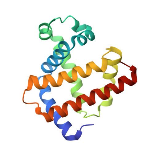
    3LR7, 3LR9

  • PubMed Abstract: 

    Exposure of a single crystal of the nitrite adduct of ferric myoglobin (Mb) at 100 K to high-intensity synchrotron X-ray radiation resulted in changes in the UV-vis spectrum that can be attributed to reduction of the ferric compound to the ferrous derivative. We employed correlated single-crystal spectroscopy with crystallography to further characterize this photoproduct. The 1.55 A resolution crystal structure of the photoproduct reveals retention of the O-binding mode for binding of nitrite to the iron center. The data are consistent with cryogenic generation and trapping, at 100 K, of a ferrous d(6) Mb(II)(ONO)* complex by photoreduction of the ferric precursor crystals using high-intensity X-ray radiation.


  • Organizational Affiliation
    • Department of Chemistry and Biochemistry, University of Oklahoma, 620 Parrington Oval, Norman, Oklahoma 73019, USA.

Macromolecule Content 

  • Total Structure Weight: 17.98 kDa 
  • Atom Count: 1,389 
  • Modeled Residue Count: 152 
  • Deposited Residue Count: 153 
  • Unique protein chains: 1

Macromolecules

Find similar proteins by:|  3D Structure
Entity ID: 1
MoleculeChains  Sequence LengthOrganismDetailsImage
Myoglobin153Equus caballusMutation(s): 0 
EC: 1.11.1 (UniProt), 1.7 (UniProt)
UniProt
Find proteins for P68082 (Equus caballus)
Explore P68082 
Go to UniProtKB:  P68082
Entity Groups
Sequence Clusters30% Identity50% Identity70% Identity90% Identity95% Identity100% Identity
UniProt GroupP68082
Sequence Annotations
Expand
Reference Sequence

Small Molecules

Ligands 3 Unique
IDChains Name / Formula / InChI Key2D Diagram3D Interactions
HEM

Query on HEM


Download:Ideal Coordinates CCD File
B [auth A]PROTOPORPHYRIN IX CONTAINING FE
C34 H32 Fe N4 O4
KABFMIBPWCXCRK-RGGAHWMASA-L
SO4

Query on SO4


Download:Ideal Coordinates CCD File
G [auth A],
H [auth A]
SULFATE ION
O4 S
QAOWNCQODCNURD-UHFFFAOYSA-L
NO2

Query on NO2


Download:Ideal Coordinates CCD File
C [auth A],
D [auth A],
E [auth A],
F [auth A]
NITRITE ION
N O2
IOVCWXUNBOPUCH-UHFFFAOYSA-M

Experimental Data & Validation

Experimental Data

  • Method: X-RAY DIFFRACTION
  • Resolution: 1.55 Å
  • R-Value Free:  0.212 (Depositor), 0.201 (DCC) 
  • R-Value Work:  0.178 (Depositor), 0.168 (DCC) 
  • R-Value Observed: 0.180 (Depositor) 
Space Group: P 1 21 1
Unit Cell:
Length ( Å )Angle ( ˚ )
a = 35.327α = 90
b = 28.691β = 105.64
c = 62.911γ = 90
Software Package:
Software NamePurpose
ADSCdata collection
PHASERphasing
PHENIXrefinement
d*TREKdata reduction
d*TREKdata scaling

Structure Validation

View Full Validation Report


Ligand Structure Quality Assessment 


Entry History 

Deposition Data

Revision History  (Full details and data files)

  • Version 1.0: 2010-07-07
    Type: Initial release
  • Version 1.1: 2011-07-13
    Changes: Version format compliance
  • Version 1.2: 2023-09-06
    Changes: Data collection, Database references, Derived calculations, Refinement description