3LQX | pdb_00003lqx

SRP ribonucleoprotein core complexed with cobalt hexammine


Experimental Data Snapshot

  • Method: X-RAY DIFFRACTION
  • Resolution: 1.93 Å
  • R-Value Free: 
    0.259 (Depositor), 0.257 (DCC) 
  • R-Value Work: 
    0.244 (Depositor), 0.235 (DCC) 

Starting Model: experimental
View more details

wwPDB Validation 3D Report Full Report

Validation slider image for 3LQX

This is version 1.5 of the entry. See complete history

Literature

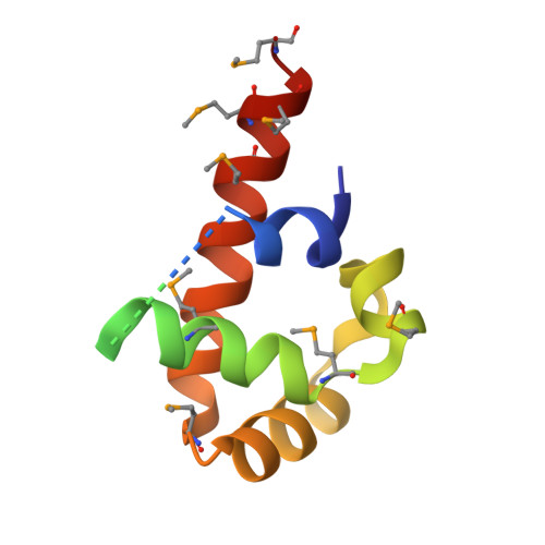
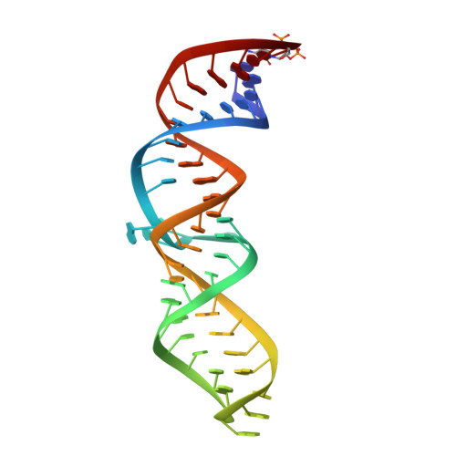
Structural and Energetic Analysis of Metal Ions Essential to SRP Signal Recognition Domain Assembly

Batey, R.T.Doudna, J.A.

(2002) Biochemistry 41: 11703-11710

  • DOI: https://doi.org/10.1021/bi026163c
  • Primary Citation Related Structures: 
    3LQX

  • PubMed Abstract: 

    The signal recognition particle (SRP) targets proteins to the endoplasmic reticulum in eukaryotes or to the inner membrane in prokaryotes by binding to hydrophobic signal sequences. Signal peptide recognition occurs within the highly conserved RNA-protein core of the SRP, underscoring the importance of this complex in SRP function. Structural analysis of the RNA and protein components of the prokaryotic SRP in the free and bound states revealed that the RNA undergoes a significant conformational change upon protein binding involving the uptake of several monovalent and divalent cations. To investigate the role of these metal ions in formation of the functional SRP complex, we used binding affinity assays and X-ray crystallography to analyze the specificity and energetic contributions of mono- and divalent metal ions bound in the RNA. Our results demonstrate that several metal ion binding sites important for RNA conformation can accommodate chemically distinct ions, often without affecting the structure of the complex. Thus, while these metal ions are highly ordered and essential for the formation and stability of the SRP complex, they behave like nonspecific metal ions.


  • Organizational Affiliation
    • Department of Molecular Biophysics and Biochemistry, Howard Hughes Medical Institute, Yale University, New Haven, CT 06520, USA.

Macromolecule Content 

  • Total Structure Weight: 30.03 kDa 
  • Atom Count: 1,741 
  • Modeled Residue Count: 118 
  • Deposited Residue Count: 154 
  • Unique protein chains: 1
  • Unique nucleic acid chains: 1

Macromolecules


Find similar proteins by:|  3D Structure
Entity ID: 1
MoleculeChains  Sequence LengthOrganismDetailsImage
Signal recognition particle protein105Escherichia coliMutation(s): 1 
EC: 3.6.5.4
UniProt
Find proteins for P0AGD7 (Escherichia coli (strain K12))
Explore P0AGD7 
Go to UniProtKB:  P0AGD7
Entity Groups
Sequence Clusters30% Identity50% Identity70% Identity90% Identity95% Identity100% Identity
UniProt GroupP0AGD7
Sequence Annotations
Expand
Reference Sequence
Find similar nucleic acids by:  (by identity cutoff) 
Entity ID: 2
MoleculeChains LengthOrganismImage
SRP RNA49N/A
Sequence Annotations
Expand
Reference Sequence

Small Molecules

Ligands 3 Unique
IDChains Name / Formula / InChI Key2D Diagram3D Interactions
NCO

Query on NCO


Download:Ideal Coordinates CCD File
F [auth B]
G [auth B]
H [auth B]
I [auth B]
J [auth B]
F [auth B],
G [auth B],
H [auth B],
I [auth B],
J [auth B],
K [auth B],
L [auth B],
M [auth B]
COBALT HEXAMMINE(III)
Co H18 N6
DYLMFCCYOUSRTK-UHFFFAOYSA-N
K

Query on K


Download:Ideal Coordinates CCD File
C [auth B],
D [auth B]
POTASSIUM ION
K
NPYPAHLBTDXSSS-UHFFFAOYSA-N
CL

Query on CL


Download:Ideal Coordinates CCD File
E [auth B]CHLORIDE ION
Cl
VEXZGXHMUGYJMC-UHFFFAOYSA-M
Modified Residues  1 Unique
IDChains TypeFormula2D DiagramParent
MSE
Query on MSE
A
L-PEPTIDE LINKINGC5 H11 N O2 SeMET

Experimental Data & Validation

Experimental Data

  • Method: X-RAY DIFFRACTION
  • Resolution: 1.93 Å
  • R-Value Free:  0.259 (Depositor), 0.257 (DCC) 
  • R-Value Work:  0.244 (Depositor), 0.235 (DCC) 
Space Group: C 1 2 1
Unit Cell:
Length ( Å )Angle ( ˚ )
a = 136.818α = 90
b = 77.765β = 94.08
c = 32.735γ = 90
Software Package:
Software NamePurpose
HKL-2000data collection
CNSrefinement
HKL-2000data reduction
HKL-2000data scaling
CNSphasing

Structure Validation

View Full Validation Report


Entry History 

Deposition Data

  • Released Date: 2010-03-02 
  • Deposition Author(s): Batey, R.T.

Revision History  (Full details and data files)

  • Version 1.0: 2010-03-02
    Type: Initial release
  • Version 1.1: 2011-07-13
    Changes: Version format compliance
  • Version 1.2: 2015-01-21
    Changes: Database references
  • Version 1.3: 2023-09-06
    Changes: Data collection, Database references, Derived calculations, Refinement description
  • Version 1.4: 2023-11-22
    Changes: Data collection
  • Version 1.5: 2024-11-20
    Changes: Structure summary