3LPT | pdb_00003lpt

HIV integrase


Experimental Data Snapshot

  • Method: X-RAY DIFFRACTION
  • Resolution: 2.00 Å
  • R-Value Free: 
    0.272 (Depositor), 0.268 (DCC) 
  • R-Value Work: 
    0.231 (Depositor), 0.229 (DCC) 
  • R-Value Observed: 
    0.233 (Depositor) 

wwPDB Validation 3D Report Full Report

Validation slider image for 3LPT

Ligand Structure Quality Assessment 


This is version 1.5 of the entry. See complete history

Literature

Rational design of small-molecule inhibitors of the LEDGF/p75-integrase interaction and HIV replication.

Christ, F.Voet, A.Marchand, A.Nicolet, S.Desimmie, B.A.Marchand, D.Bardiot, D.Van der Veken, N.J.Van Remoortel, B.Strelkov, S.V.De Maeyer, M.Chaltin, P.Debyser, Z.

(2010) Nat Chem Biol 6: 442-448

  • DOI: https://doi.org/10.1038/nchembio.370
  • Primary Citation Related Structures: 
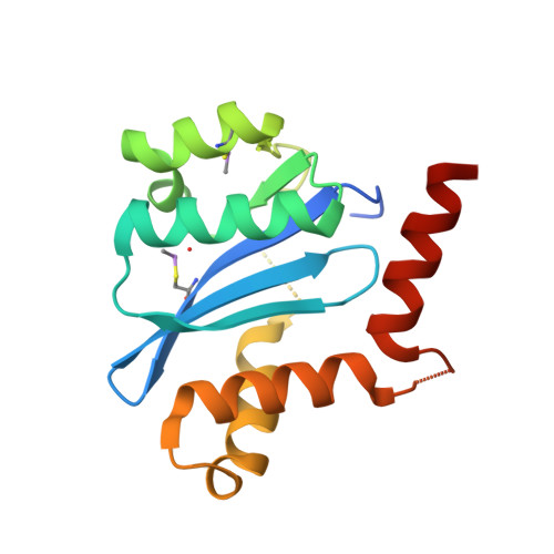
    3LPT, 3LPU

  • PubMed Abstract: 

    Lens epithelium-derived growth factor (LEDGF/p75) is a cellular cofactor of HIV-1 integrase that promotes viral integration by tethering the preintegration complex to the chromatin. By virtue of its crucial role in the early steps of HIV replication, the interaction between LEDGF/p75 and integrase represents an attractive target for antiviral therapy. We have rationally designed a series of 2-(quinolin-3-yl)acetic acid derivatives (LEDGINs) that act as potent inhibitors of the LEDGF/p75-integrase interaction and HIV-1 replication at submicromolar concentration by blocking the integration step. A 1.84-A resolution crystal structure corroborates the binding of the inhibitor in the LEDGF/p75-binding pocket of integrase. Together with the lack of cross-resistance with two clinical integrase inhibitors, these findings define the 2-(quinolin-3-yl)acetic acid derivatives as the first genuine allosteric HIV-1 integrase inhibitors. Our work demonstrates the feasibility of rational design of small molecules inhibiting the protein-protein interaction between a viral protein and a cellular host factor.


  • Organizational Affiliation
    • Laboratory for Molecular Virology and Gene Therapy, Division of Molecular Medicine, Katholieke Universiteit Leuven (KULeuven), Leuven, Belgium.

Macromolecule Content 

  • Total Structure Weight: 19.76 kDa 
  • Atom Count: 1,324 
  • Modeled Residue Count: 144 
  • Deposited Residue Count: 166 
  • Unique protein chains: 1

Macromolecules

Find similar proteins by:|  3D Structure
Entity ID: 1
MoleculeChains  Sequence LengthOrganismDetailsImage
Integrase166Human immunodeficiency virus 1Mutation(s): 1 
UniProt
Find proteins for P12497 (Human immunodeficiency virus type 1 group M subtype B (isolate NY5))
Explore P12497 
Go to UniProtKB:  P12497
Entity Groups
Sequence Clusters30% Identity50% Identity70% Identity90% Identity95% Identity100% Identity
UniProt GroupP12497
Sequence Annotations
Expand
Reference Sequence

Small Molecules

Ligands 6 Unique
IDChains Name / Formula / InChI Key2D Diagram3D Interactions
723

Query on 723


Download:Ideal Coordinates CCD File
B [auth A](6-chloro-2-oxo-4-phenyl-1,2-dihydroquinolin-3-yl)acetic acid
C17 H12 Cl N O3
SHDUUQWYFNJIAG-UHFFFAOYSA-N
P03

Query on P03


Download:Ideal Coordinates CCD File
F [auth A]2-[3-[3-(2-hydroxyethoxy)propoxy]propoxy]ethanol
C10 H22 O5
GNAZJSIZGCDIQL-UHFFFAOYSA-N
PEG

Query on PEG


Download:Ideal Coordinates CCD File
C [auth A],
D [auth A],
E [auth A],
G [auth A]
DI(HYDROXYETHYL)ETHER
C4 H10 O3
MTHSVFCYNBDYFN-UHFFFAOYSA-N
SO4

Query on SO4


Download:Ideal Coordinates CCD File
H [auth A],
I [auth A],
J [auth A]
SULFATE ION
O4 S
QAOWNCQODCNURD-UHFFFAOYSA-L
CA

Query on CA


Download:Ideal Coordinates CCD File
K [auth A]CALCIUM ION
Ca
BHPQYMZQTOCNFJ-UHFFFAOYSA-N
CL

Query on CL


Download:Ideal Coordinates CCD File
L [auth A]CHLORIDE ION
Cl
VEXZGXHMUGYJMC-UHFFFAOYSA-M
Modified Residues  1 Unique
IDChains TypeFormula2D DiagramParent
CAF
Query on CAF
A
L-PEPTIDE LINKINGC5 H12 As N O3 SCYS
Binding Affinity Annotations 
IDSourceBinding Affinity
723 BindingDB:  3LPT IC50: 1.17e+4 (nM) from 1 assay(s)

Experimental Data & Validation

Experimental Data

  • Method: X-RAY DIFFRACTION
  • Resolution: 2.00 Å
  • R-Value Free:  0.272 (Depositor), 0.268 (DCC) 
  • R-Value Work:  0.231 (Depositor), 0.229 (DCC) 
  • R-Value Observed: 0.233 (Depositor) 
Space Group: P 31 2 1
Unit Cell:
Length ( Å )Angle ( ˚ )
a = 72α = 90
b = 72β = 90
c = 65.49γ = 120
Software Package:
Software NamePurpose
MAR345dtbdata collection
REFMACrefinement
MOSFLMdata reduction
SCALAdata scaling

Structure Validation

View Full Validation Report


Ligand Structure Quality Assessment 


Entry History 

Deposition Data

Revision History  (Full details and data files)

  • Version 1.0: 2010-05-12
    Type: Initial release
  • Version 1.1: 2011-07-13
    Changes: Version format compliance
  • Version 1.2: 2015-12-09
    Changes: Database references
  • Version 1.3: 2017-11-01
    Changes: Refinement description
  • Version 1.4: 2021-11-10
    Changes: Database references, Derived calculations
  • Version 1.5: 2024-10-30
    Changes: Data collection, Structure summary