3LO2 | pdb_00003lo2

Crystal structure of human alpha-defensin 1 (Y21A mutant)


Experimental Data Snapshot

  • Method: X-RAY DIFFRACTION
  • Resolution: 1.56 Å
  • R-Value Free: 
    0.200 (Depositor), 0.210 (DCC) 
  • R-Value Work: 
    0.173 (Depositor), 0.185 (DCC) 
  • R-Value Observed: 
    0.175 (Depositor) 

Starting Model: experimental
View more details

wwPDB Validation 3D Report Full Report

Validation slider image for 3LO2

This is version 1.4 of the entry. See complete history

Literature

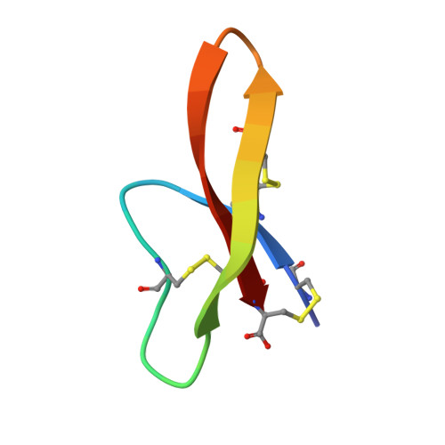
Trp-26 imparts functional versatility to human alpha-defensin HNP1.

Wei, G.Pazgier, M.de Leeuw, E.Rajabi, M.Li, J.Zou, G.Jung, G.Yuan, W.Lu, W.Y.Lehrer, R.I.Lu, W.

(2010) J Biological Chem 285: 16275-16285

  • DOI: https://doi.org/10.1074/jbc.M110.102749
  • Primary Citation Related Structures: 
    3H6C, 3LO1, 3LO2, 3LO4, 3LO6, 3LO9, 3LOE, 3LVX

  • PubMed Abstract: 

    We performed a comprehensive alanine scan of human alpha-defensin HNP1 and tested the ability of the resulting analogs to kill Staphylococcus aureus, inhibit anthrax lethal factor, and bind human immunodeficiency virus-1 gp120. By far, the most deleterious mutation for all of these functions was W26A. The activities lost by W26A-HNP1 were restored progressively by replacing W26 with non-coded, straight-chain aliphatic amino acids of increasing chain length. The hydrophobicity of residue 26 also correlated with the ability of the analogs to bind immobilized wild type HNP1 and to undergo further self-association. Thus, the hydrophobicity of residue 26 is not only a key determinant of the direct interactions of HNP1 with target molecules, but it also governs the ability of this peptide to form dimers and more complex quaternary structures at micromolar concentrations. Although all defensin peptides are cationic, their amphipathicity is at least as important as their positive charge in enabling them to participate in innate host defense.


  • Organizational Affiliation
    • Institute of Human Virology, University of Maryland School of Medicine, Baltimore, Maryland 21201, USA.

Macromolecule Content 

  • Total Structure Weight: 7.17 kDa 
  • Atom Count: 563 
  • Modeled Residue Count: 60 
  • Deposited Residue Count: 60 
  • Unique protein chains: 1

Macromolecules

Find similar proteins by:|  3D Structure
Entity ID: 1
MoleculeChains  Sequence LengthOrganismDetailsImage
Neutrophil defensin 1
A, B
30N/AMutation(s): 1 
UniProt & NIH Common Fund Data Resources
Find proteins for P59665 (Homo sapiens)
Explore P59665 
Go to UniProtKB:  P59665
Entity Groups
Sequence Clusters30% Identity50% Identity70% Identity90% Identity95% Identity100% Identity
UniProt GroupP59665
Sequence Annotations
Expand
Reference Sequence

Experimental Data & Validation

Experimental Data

  • Method: X-RAY DIFFRACTION
  • Resolution: 1.56 Å
  • R-Value Free:  0.200 (Depositor), 0.210 (DCC) 
  • R-Value Work:  0.173 (Depositor), 0.185 (DCC) 
  • R-Value Observed: 0.175 (Depositor) 
Space Group: P 21 21 2
Unit Cell:
Length ( Å )Angle ( ˚ )
a = 43.675α = 90
b = 29.882β = 90
c = 41.552γ = 90
Software Package:
Software NamePurpose
REFMACrefinement
PDB_EXTRACTdata extraction
HKL-2000data collection
DENZOdata reduction
SCALEPACKdata scaling
PHASERphasing

Structure Validation

View Full Validation Report


Entry History 

Deposition Data

Revision History  (Full details and data files)

  • Version 1.0: 2010-03-09
    Type: Initial release
  • Version 1.1: 2011-07-13
    Changes: Refinement description, Version format compliance
  • Version 1.2: 2021-10-13
    Changes: Database references, Derived calculations
  • Version 1.3: 2023-09-06
    Changes: Data collection, Refinement description
  • Version 1.4: 2024-11-27
    Changes: Structure summary