3LNV | pdb_00003lnv

The crystal structure of fatty acyl-adenylate ligase from L. pneumophila in complex with acyl adenylate and pyrophosphate


Experimental Data Snapshot

  • Method: X-RAY DIFFRACTION
  • Resolution: 2.00 Å
  • R-Value Free: 
    0.244 (Depositor), 0.192 (DCC) 
  • R-Value Work: 
    0.193 (Depositor), 0.197 (DCC) 
  • R-Value Observed: 
    0.196 (Depositor) 

Starting Model: experimental
View more details

wwPDB Validation 3D Report Full Report

Validation slider image for 3LNV

Ligand Structure Quality Assessment 


This is version 1.5 of the entry. See complete history

Literature

Structural and Functional Studies of Fatty Acyl Adenylate Ligases from E. coli and L. pneumophila.

Zhang, Z.Zhou, R.Sauder, J.M.Tonge, P.J.Burley, S.K.Swaminathan, S.

(2011) J Mol Biology 406: 313-324

  • DOI: https://doi.org/10.1016/j.jmb.2010.12.011
  • Primary Citation Related Structures: 
    3KXW, 3LNV, 3PBK

  • PubMed Abstract: 

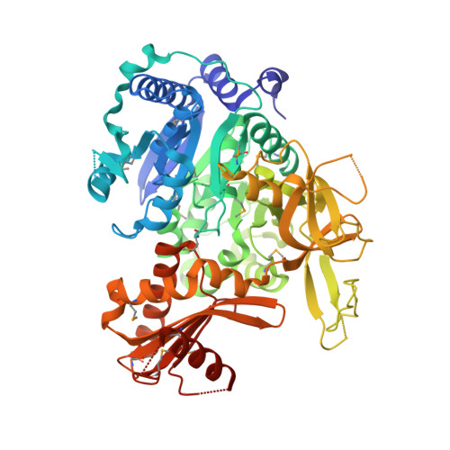
    Fatty acyl-AMP ligase (FAAL) is a new member of a family of adenylate-forming enzymes that were recently discovered in Mycobacterium tuberculosis. They are similar in sequence to fatty acyl-coenzyme A (CoA) ligases (FACLs). However, while FACLs perform a two-step catalytic reaction, AMP ligation followed by CoA ligation using ATP and CoA as cofactors, FAALs produce only the acyl adenylate and are unable to perform the second step. We report X-ray crystal structures of full-length FAAL from Escherichia coli (EcFAAL) and FAAL from Legionella pneumophila (LpFAAL) bound to acyl adenylate, determined at resolution limits of 3.0 and 1.85 Å, respectively. The structures share a larger N-terminal domain and a smaller C-terminal domain, which together resemble the previously determined structures of FAAL and FACL proteins. Our two structures occur in quite different conformations. EcFAAL adopts the adenylate-forming conformation typical of FACLs, whereas LpFAAL exhibits a unique intermediate conformation. Both EcFAAL and LpFAAL have insertion motifs that distinguish them from the FACLs. Structures of EcFAAL and LpFAAL reveal detailed interactions between this insertion motif and the interdomain hinge region and with the C-terminal domain. We suggest that the insertion motifs support sufficient interdomain motions to allow substrate binding and product release during acyl adenylate formation, but they preclude CoA binding, thereby preventing CoA ligation.


  • Organizational Affiliation
    • Biology Department, Brookhaven National Laboratory, Upton, NY 11973, USA.

Macromolecule Content 

  • Total Structure Weight: 68.57 kDa 
  • Atom Count: 4,957 
  • Modeled Residue Count: 566 
  • Deposited Residue Count: 590 
  • Unique protein chains: 1

Macromolecules

Find similar proteins by:|  3D Structure
Entity ID: 1
MoleculeChains  Sequence LengthOrganismDetailsImage
Saframycin Mx1 synthetase B590Legionella pneumophila subsp. pneumophila str. Philadelphia 1Mutation(s): 0 
Gene Names: 11191l2lpg2229
UniProt
Find proteins for Q5ZTD3 (Legionella pneumophila subsp. pneumophila (strain Philadelphia 1 / ATCC 33152 / DSM 7513))
Explore Q5ZTD3 
Go to UniProtKB:  Q5ZTD3
Entity Groups
Sequence Clusters30% Identity50% Identity70% Identity90% Identity95% Identity100% Identity
UniProt GroupQ5ZTD3
Sequence Annotations
Expand
Reference Sequence

Small Molecules

Ligands 2 Unique
IDChains Name / Formula / InChI Key2D Diagram3D Interactions
1ZZ

Query on 1ZZ


Download:Ideal Coordinates CCD File
B [auth A]5'-O-[(S)-(dodecanoyloxy)(hydroxy)phosphoryl]adenosine
C22 H36 N5 O8 P
IKBWVSPLSBIYSK-CIVUBGFFSA-N
POP

Query on POP


Download:Ideal Coordinates CCD File
C [auth A]PYROPHOSPHATE 2-
H2 O7 P2
XPPKVPWEQAFLFU-UHFFFAOYSA-L
Modified Residues  1 Unique
IDChains TypeFormula2D DiagramParent
MSE
Query on MSE
A
L-PEPTIDE LINKINGC5 H11 N O2 SeMET

Experimental Data & Validation

Experimental Data

  • Method: X-RAY DIFFRACTION
  • Resolution: 2.00 Å
  • R-Value Free:  0.244 (Depositor), 0.192 (DCC) 
  • R-Value Work:  0.193 (Depositor), 0.197 (DCC) 
  • R-Value Observed: 0.196 (Depositor) 
Space Group: P 21 21 21
Unit Cell:
Length ( Å )Angle ( ˚ )
a = 70.193α = 90
b = 78.243β = 90
c = 108.395γ = 90
Software Package:
Software NamePurpose
CBASSdata collection
PHASERphasing
PHENIXrefinement
MOSFLMdata reduction
SCALAdata scaling

Structure Validation

View Full Validation Report


Ligand Structure Quality Assessment 


Entry History 

Deposition Data

Revision History  (Full details and data files)

  • Version 1.0: 2010-04-07
    Type: Initial release
  • Version 1.1: 2011-07-13
    Changes: Version format compliance
  • Version 1.2: 2021-02-10
    Changes: Database references, Derived calculations, Structure summary
  • Version 1.3: 2023-09-06
    Changes: Data collection, Database references, Refinement description
  • Version 1.4: 2023-11-22
    Changes: Data collection
  • Version 1.5: 2024-11-06
    Changes: Structure summary