3LLN | pdb_00003lln

Comparison between the orthorhombic an tetragonal form of the heptamer sequence d(GCG(xT)GCG)/d(CGCACGC).


Experimental Data Snapshot

  • Method: X-RAY DIFFRACTION
  • Resolution: 3.00 Å
  • R-Value Work: 
    0.226 (Depositor), 0.242 (DCC) 
  • R-Value Observed: 
    0.226 (Depositor) 

Starting Model: experimental
View more details

wwPDB Validation 3D Report Full Report

Validation slider image for 3LLN

This is version 1.2 of the entry. See complete history

Literature

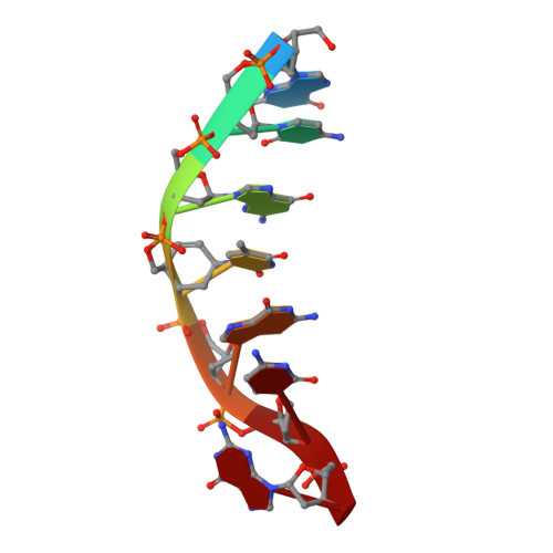
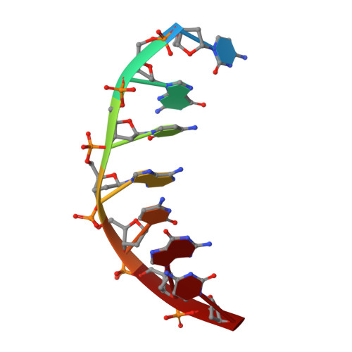
Comparison between the orthorhombic and tetragonal forms of the heptamer sequence d[GCG(xT)GCG]/d(CGCACGC)

Robeyns, K.Herdewijn, P.Van Meervelt, L.

(2010) Acta Crystallogr Sect F Struct Biol Cryst Commun 66: 1028-1031

  • DOI: https://doi.org/10.1107/S1744309110031696
  • Primary Citation Related Structures: 
    3LLN

  • PubMed Abstract: 

    Cyclohexene nucleic acid (CeNA) building blocks can be introduced into natural DNA sequences without a large conformational influence because of the ability of the six-membered sugar ring to mimic both the C2'-endo and C3'-endo conformations of the naturally occurring ribofuranose sugar ring. The non-self-complementary DNA sequence d[GCG(xT)GCG]/d(CGCACGC) with one incorporated CeNA (xT) moiety crystallizes in two forms: orthorhombic and tetragonal. The tetragonal form, which diffracts to 3 A resolution, is a kinetically stable polymorph of the orthorhombic form [Robeyns et al. (2010), Artificial DNA, 1, 1-7], which diffracts to 1.17 A resolution and is the thermodynamically stable form of the CeNA-incorporated duplex. Here, the two structures are compared, with special emphasis on the differences in crystal packing and the irreversible conversion of the kinetic form into the high-resolution diffracting thermodynamic form.


  • Organizational Affiliation
    • Department of Chemistry, Biomolecular Architecture and BioMacS, Katholieke Universiteit Leuven, Belgium.

Macromolecule Content 

  • Total Structure Weight: 4.41 kDa 
  • Atom Count: 298 
  • Modeled Residue Count: 14 
  • Deposited Residue Count: 14 
  • Unique nucleic acid chains: 2

Macromolecules

Find similar nucleic acids by:  Sequence
Entity ID: 1
MoleculeChains LengthOrganismImage
DNA (5'-D(*GP*CP*GP*(XTR)P*GP*CP*G)-3')7N/A
Sequence Annotations
Expand
Reference Sequence
Find similar nucleic acids by:  Sequence
Entity ID: 2
MoleculeChains LengthOrganismImage
DNA (5'-D(*CP*GP*CP*AP*CP*GP*C)-3')7N/A
Sequence Annotations
Expand
Reference Sequence

Small Molecules

Ligands 1 Unique
IDChains Name / Formula / InChI Key2D Diagram3D Interactions
NCO

Query on NCO


Download:Ideal Coordinates CCD File
C [auth A]COBALT HEXAMMINE(III)
Co H18 N6
DYLMFCCYOUSRTK-UHFFFAOYSA-N

Experimental Data & Validation

Experimental Data

  • Method: X-RAY DIFFRACTION
  • Resolution: 3.00 Å
  • R-Value Work:  0.226 (Depositor), 0.242 (DCC) 
  • R-Value Observed: 0.226 (Depositor) 
Space Group: P 41 21 2
Unit Cell:
Length ( Å )Angle ( ˚ )
a = 25.482α = 90
b = 25.482β = 90
c = 81.147γ = 90
Software Package:
Software NamePurpose
MAR345dtbdata collection
PHASERphasing
REFMACrefinement
MOSFLMdata reduction
SCALAdata scaling

Structure Validation

View Full Validation Report


Entry History 

Deposition Data

Revision History  (Full details and data files)

  • Version 1.0: 2010-08-18
    Type: Initial release
  • Version 1.1: 2011-07-13
    Changes: Version format compliance
  • Version 1.2: 2023-09-06
    Changes: Data collection, Database references, Derived calculations, Refinement description