3LLI | pdb_00003lli

Sulfhydryl Oxidase Fragment of Human QSOX1


Experimental Data Snapshot

  • Method: X-RAY DIFFRACTION
  • Resolution: 2.05 Å
  • R-Value Free: 
    0.325 (Depositor), 0.323 (DCC) 
  • R-Value Work: 
    0.275 (Depositor), 0.275 (DCC) 

wwPDB Validation 3D Report Full Report

Validation slider image for 3LLI

Ligand Structure Quality Assessment 


This is version 1.2 of the entry. See complete history

Literature

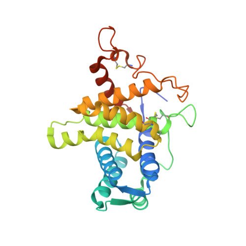
QSOX contains a pseudo-dimer of functional and degenerate sulfhydryl oxidase domains.

Alon, A.Heckler, E.J.Thorpe, C.Fass, D.

(2010) FEBS Lett 584: 1521-1525

  • DOI: https://doi.org/10.1016/j.febslet.2010.03.001
  • Primary Citation Related Structures: 
    3LLI, 3LLK

  • PubMed Abstract: 

    Quiescin sulfhydryl oxidase (QSOX) catalyzes formation of disulfide bonds between cysteine residues in substrate proteins. Human QSOX1 is a multi-domain, monomeric enzyme containing a module related to the single-domain sulfhydryl oxidases of the Erv family. A partial QSOX1 crystal structure reveals a single-chain pseudo-dimer mimicking the quaternary structure of Erv enzymes. However, one pseudo-dimer "subunit" has lost its cofactor and catalytic activity. In QSOX evolution, a further concatenation to a member of the protein disulfide isomerase family resulted in an enzyme capable of both disulfide formation and efficient transfer to substrate proteins.


  • Organizational Affiliation
    • Department of Structural Biology, Weizmann Institute of Science, Rehovot, Israel.

Macromolecule Content 

  • Total Structure Weight: 30.58 kDa 
  • Atom Count: 2,178 
  • Modeled Residue Count: 251 
  • Deposited Residue Count: 261 
  • Unique protein chains: 1

Macromolecules

Find similar proteins by:|  3D Structure
Entity ID: 1
MoleculeChains  Sequence LengthOrganismDetailsImage
Sulfhydryl oxidase 1261Homo sapiensMutation(s): 0 
Gene Names: QSCN6QSOX1UNQ2520/PRO6013
EC: 1.8.3.2
UniProt & NIH Common Fund Data Resources
Find proteins for O00391 (Homo sapiens)
Explore O00391 
Go to UniProtKB:  O00391
PHAROS:  O00391
GTEx:  ENSG00000116260 
Entity Groups
Sequence Clusters30% Identity50% Identity70% Identity90% Identity95% Identity100% Identity
UniProt GroupO00391
Sequence Annotations
Expand
Reference Sequence

Small Molecules

Ligands 1 Unique
IDChains Name / Formula / InChI Key2D Diagram3D Interactions
FAD

Query on FAD


Download:Ideal Coordinates CCD File
B [auth A]FLAVIN-ADENINE DINUCLEOTIDE
C27 H33 N9 O15 P2
VWWQXMAJTJZDQX-UYBVJOGSSA-N

Experimental Data & Validation

Experimental Data

  • Method: X-RAY DIFFRACTION
  • Resolution: 2.05 Å
  • R-Value Free:  0.325 (Depositor), 0.323 (DCC) 
  • R-Value Work:  0.275 (Depositor), 0.275 (DCC) 
Space Group: P 64 2 2
Unit Cell:
Length ( Å )Angle ( ˚ )
a = 85.606α = 90
b = 85.606β = 90
c = 125.706γ = 120
Software Package:
Software NamePurpose
SOLVEphasing
CNSrefinement
DENZOdata reduction
SCALEPACKdata scaling

Structure Validation

View Full Validation Report


Ligand Structure Quality Assessment 


Entry History 

Deposition Data

Revision History  (Full details and data files)

  • Version 1.0: 2010-03-23
    Type: Initial release
  • Version 1.1: 2011-07-13
    Changes: Version format compliance
  • Version 1.2: 2024-11-27
    Changes: Data collection, Database references, Derived calculations, Structure summary