3LJT | pdb_00003ljt

Crystal Structure of the Catalytic Domain of ADAMTS-5 in Complex with an Amino-2-indanol compound


Experimental Data Snapshot

  • Method: X-RAY DIFFRACTION
  • Resolution: 1.60 Å
  • R-Value Free: 
    0.198 (Depositor), 0.205 (DCC) 
  • R-Value Work: 
    0.159 (Depositor), 0.166 (DCC) 
  • R-Value Observed: 
    0.161 (Depositor) 

Starting Model: experimental
View more details

wwPDB Validation 3D Report Full Report

Validation slider image for 3LJT

Ligand Structure Quality Assessment 


This is version 1.5 of the entry. See complete history

Literature

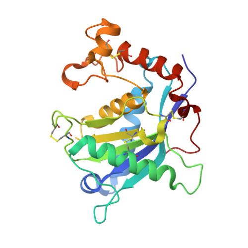
Structure analysis reveals the flexibility of the ADAMTS-5 active site.

Shieh, H.S.Tomasselli, A.G.Mathis, K.J.Schnute, M.E.Woodard, S.S.Caspers, N.Williams, J.M.Kiefer, J.R.Munie, G.Wittwer, A.Malfait, A.M.Tortorella, M.D.

(2011) Protein Sci 20: 735-744

  • DOI: https://doi.org/10.1002/pro.606
  • Primary Citation Related Structures: 
    3LJT, 3LJZ

  • PubMed Abstract: 

    A ((1S,2R)-2-hydroxy-2,3-dihydro-1H-inden-1-yl) succinamide derivative (here referred to as Compound 12) shows significant activity toward many matrix metalloproteinases (MMPs), including MMP-2, MMP-8, MMP-9, and MMP-13. Modeling studies had predicted that this compound would not bind to ADAMTS-5 (a disintegrin and metalloproteinase with thrombospondin motifs-5) due to its shallow S1' pocket. However, inhibition analysis revealed it to be a nanomolar inhibitor of both ADAMTS-4 and -5. The observed inconsistency was explained by analysis of crystallographic structures, which showed that Compound 12 in complex with the catalytic domain of ADAMTS-5 (cataTS5) exhibits an unusual conformation in the S1' pocket of the protein. This first demonstration that cataTS5 can undergo an induced conformational change in its active site pocket by a molecule like Compound 12 should enable the design of new aggrecanase inhibitors with better potency and selectivity profiles.


  • Organizational Affiliation
    • Pfizer Global Research and Development, St. Louis, Missouri 63017, USA. shiehouse@sbcglobal.net

Macromolecule Content 

  • Total Structure Weight: 24.83 kDa 
  • Atom Count: 2,045 
  • Modeled Residue Count: 218 
  • Deposited Residue Count: 218 
  • Unique protein chains: 1

Macromolecules

Find similar proteins by:|  3D Structure
Entity ID: 1
MoleculeChains  Sequence LengthOrganismDetailsImage
A disintegrin and metalloproteinase with thrombospondin motifs 5218Homo sapiensMutation(s): 1 
Gene Names: ADAMTS-5ADAMTS11ADAMTS5ADMP2
EC: 3.4.24
UniProt & NIH Common Fund Data Resources
Find proteins for Q9UNA0 (Homo sapiens)
Explore Q9UNA0 
Go to UniProtKB:  Q9UNA0
PHAROS:  Q9UNA0
GTEx:  ENSG00000154736 
Entity Groups
Sequence Clusters30% Identity50% Identity70% Identity90% Identity95% Identity100% Identity
UniProt GroupQ9UNA0
Sequence Annotations
Expand
Reference Sequence

Small Molecules

Ligands 4 Unique
IDChains Name / Formula / InChI Key2D Diagram3D Interactions
LA3

Query on LA3


Download:Ideal Coordinates CCD File
B [auth A](2R)-2-[4-(1,3-benzodioxol-5-yl)benzyl]-N~4~-hydroxy-N~1~-[(1S,2R)-2-hydroxy-2,3-dihydro-1H-inden-1-yl]butanediamide
C27 H26 N2 O6
MJTVUROZTDIMFT-MZEQIWSPSA-N
ZN

Query on ZN


Download:Ideal Coordinates CCD File
D [auth A]ZINC ION
Zn
PTFCDOFLOPIGGS-UHFFFAOYSA-N
EDO

Query on EDO


Download:Ideal Coordinates CCD File
C [auth A]1,2-ETHANEDIOL
C2 H6 O2
LYCAIKOWRPUZTN-UHFFFAOYSA-N
CA

Query on CA


Download:Ideal Coordinates CCD File
E [auth A],
F [auth A],
G [auth A]
CALCIUM ION
Ca
BHPQYMZQTOCNFJ-UHFFFAOYSA-N

Experimental Data & Validation

Experimental Data

  • Method: X-RAY DIFFRACTION
  • Resolution: 1.60 Å
  • R-Value Free:  0.198 (Depositor), 0.205 (DCC) 
  • R-Value Work:  0.159 (Depositor), 0.166 (DCC) 
  • R-Value Observed: 0.161 (Depositor) 
Space Group: P 21 21 21
Unit Cell:
Length ( Å )Angle ( ˚ )
a = 51.398α = 90
b = 44.518β = 90
c = 76.251γ = 90
Software Package:
Software NamePurpose
HKL-2000data collection
REFMACrefinement
HKL-2000data reduction
HKL-2000data scaling

Structure Validation

View Full Validation Report


Ligand Structure Quality Assessment 


Entry History 

Deposition Data

Revision History  (Full details and data files)

  • Version 1.0: 2010-03-31
    Type: Initial release
  • Version 1.1: 2011-07-13
    Changes: Version format compliance
  • Version 1.2: 2017-11-01
    Changes: Refinement description
  • Version 1.3: 2021-10-13
    Changes: Database references, Derived calculations
  • Version 1.4: 2023-09-06
    Changes: Data collection, Refinement description
  • Version 1.5: 2024-11-20
    Changes: Structure summary