3LG4 | pdb_00003lg4

Staphylococcus aureus V31Y, F92I mutant dihydrofolate reductase complexed with NADPH and 5-[(3S)-3-(5-methoxy-2',6'-dimethylbiphenyl-3-yl)but-1-yn-1-yl]-6-methylpyrimidine-2,4-diamine


Experimental Data Snapshot

  • Method: X-RAY DIFFRACTION
  • Resolution: 3.15 Å
  • R-Value Free: 
    0.292 (Depositor), 0.290 (DCC) 
  • R-Value Work: 
    0.260 (Depositor), 0.258 (DCC) 
  • R-Value Observed: 
    0.261 (Depositor) 

Starting Model: experimental
View more details

wwPDB Validation 3D Report Full Report

Validation slider image for 3LG4

Ligand Structure Quality Assessment 


This is version 1.3 of the entry. See complete history

Literature

Predicting resistance mutations using protein design algorithms.

Frey, K.M.Georgiev, I.Donald, B.R.Anderson, A.C.

(2010) Proc Natl Acad Sci U S A 107: 13707-13712

  • DOI: https://doi.org/10.1073/pnas.1002162107
  • Primary Citation Related Structures: 
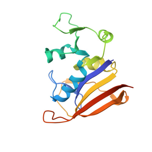
    3F0Q, 3LG4

  • PubMed Abstract: 

    Drug resistance resulting from mutations to the target is an unfortunate common phenomenon that limits the lifetime of many of the most successful drugs. In contrast to the investigation of mutations after clinical exposure, it would be powerful to be able to incorporate strategies early in the development process to predict and overcome the effects of possible resistance mutations. Here we present a unique prospective application of an ensemble-based protein design algorithm, K*, to predict potential resistance mutations in dihydrofolate reductase from Staphylococcus aureus using positive design to maintain catalytic function and negative design to interfere with binding of a lead inhibitor. Enzyme inhibition assays show that three of the four highly-ranked predicted mutants are active yet display lower affinity (18-, 9-, and 13-fold) for the inhibitor. A crystal structure of the top-ranked mutant enzyme validates the predicted conformations of the mutated residues and the structural basis of the loss of potency. The use of protein design algorithms to predict resistance mutations could be incorporated in a lead design strategy against any target that is susceptible to mutational resistance.


  • Organizational Affiliation
    • Department of Pharmaceutical Sciences, University of Connecticut, Storrs, CT 06269, USA.

Macromolecule Content 

  • Total Structure Weight: 41.31 kDa 
  • Atom Count: 2,702 
  • Modeled Residue Count: 314 
  • Deposited Residue Count: 336 
  • Unique protein chains: 1

Macromolecules

Find similar proteins by:|  3D Structure
Entity ID: 1
MoleculeChains  Sequence LengthOrganismDetailsImage
Dihydrofolate reductase
A, B
168Staphylococcus aureusMutation(s): 2 
Gene Names: dfrAdfrBfolA
EC: 1.5.1.3
UniProt
Find proteins for P0A017 (Staphylococcus aureus)
Explore P0A017 
Go to UniProtKB:  P0A017
Entity Groups
Sequence Clusters30% Identity50% Identity70% Identity90% Identity95% Identity100% Identity
UniProt GroupP0A017
Sequence Annotations
Expand
Reference Sequence

Experimental Data & Validation

Experimental Data

  • Method: X-RAY DIFFRACTION
  • Resolution: 3.15 Å
  • R-Value Free:  0.292 (Depositor), 0.290 (DCC) 
  • R-Value Work:  0.260 (Depositor), 0.258 (DCC) 
  • R-Value Observed: 0.261 (Depositor) 
Space Group: P 61
Unit Cell:
Length ( Å )Angle ( ˚ )
a = 88.752α = 90
b = 88.752β = 90
c = 103.167γ = 120
Software Package:
Software NamePurpose
REFMACrefinement
PDB_EXTRACTdata extraction
CBASSdata collection
HKL-2000data reduction
SCALEPACKdata scaling
Cootmodel building

Structure Validation

View Full Validation Report


Ligand Structure Quality Assessment 


Entry History 

Deposition Data

Revision History  (Full details and data files)

  • Version 1.0: 2010-07-28
    Type: Initial release
  • Version 1.1: 2011-07-13
    Changes: Version format compliance
  • Version 1.2: 2013-11-27
    Changes: Non-polymer description
  • Version 1.3: 2023-09-06
    Changes: Data collection, Database references, Derived calculations, Refinement description