3LFB | pdb_00003lfb

Human p38 MAP Kinase in Complex with RL98


Experimental Data Snapshot

  • Method: X-RAY DIFFRACTION
  • Resolution: 2.60 Å
  • R-Value Free: 
    0.320 (Depositor), 0.310 (DCC) 
  • R-Value Work: 
    0.230 (Depositor), 0.230 (DCC) 
  • R-Value Observed: 
    0.236 (Depositor) 

Starting Model: experimental
View more details

wwPDB Validation   3D Report Full Report


Ligand Structure Quality Assessment 

Created with Raphaël 2.3.0Worse 01 BetterLigand structure goodness of fit to experimental dataBest fitted Z87Click on this verticalbar to view detailsBest fitted BOGClick on this verticalbar to view details

This is version 1.4 of the entry. See complete history


Literature

Development of novel thiazole-urea compounds which stabalize the inactive conformation of p38 alpha

Getlik, M.Gruetter, C.Simard, J.R.Van Otterlo, W.Robubi, A.Aust, B.Rauh, D.

To be published.

Macromolecules
Find similar proteins by:  (by identity cutoff)  |  3D Structure
Entity ID: 1
MoleculeChains Sequence LengthOrganismDetailsImage
Mitogen-activated protein kinase 14360Homo sapiensMutation(s): 4 
Gene Names: MAPK14CSBPCSBP1CSBP2CSPB1MXI2
EC: 2.7.11.24
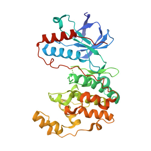
UniProt & NIH Common Fund Data Resources
Find proteins for Q16539 (Homo sapiens)
Explore Q16539 
Go to UniProtKB:  Q16539
PHAROS:  Q16539
GTEx:  ENSG00000112062 
Entity Groups  
Sequence Clusters30% Identity50% Identity70% Identity90% Identity95% Identity100% Identity
UniProt GroupQ16539
Sequence Annotations
Expand
  • Reference Sequence
Small Molecules
Ligands 2 Unique
IDChains Name / Formula / InChI Key2D Diagram3D Interactions
Z87
Query on Z87

Download Ideal Coordinates CCD File 
B [auth A]1-[3-tert-butyl-1-(4-methylphenyl)-1H-pyrazol-5-yl]-3-(1,3-thiazol-2-yl)urea
C18 H21 N5 O S
YHFISDMDEBLKPE-UHFFFAOYSA-N
BOG
Query on BOG

Download Ideal Coordinates CCD File 
C [auth A]octyl beta-D-glucopyranoside
C14 H28 O6
HEGSGKPQLMEBJL-RKQHYHRCSA-N
Binding Affinity Annotations 
IDSourceBinding Affinity
Z87 BindingDB:  3LFB Kd: 990 (nM) from 1 assay(s)
Experimental Data & Validation

Experimental Data

  • Method: X-RAY DIFFRACTION
  • Resolution: 2.60 Å
  • R-Value Free:  0.320 (Depositor), 0.310 (DCC) 
  • R-Value Work:  0.230 (Depositor), 0.230 (DCC) 
  • R-Value Observed: 0.236 (Depositor) 
Space Group: P 21 21 21
Unit Cell:
Length ( Å )Angle ( ˚ )
a = 67.12α = 90
b = 69.89β = 90
c = 74.43γ = 90
Software Package:
Software NamePurpose
XSCALEdata scaling
PHASERphasing
REFMACrefinement
PDB_EXTRACTdata extraction
XDSdata scaling
XDSdata reduction

Structure Validation

View Full Validation Report


Ligand Structure Quality Assessment 

Created with Raphaël 2.3.0Worse 01 BetterLigand structure goodness of fit to experimental dataBest fitted Z87Click on this verticalbar to view detailsBest fitted BOGClick on this verticalbar to view details

Entry History 

Deposition Data

Revision History  (Full details and data files)

  • Version 1.0: 2011-04-20
    Type: Initial release
  • Version 1.1: 2011-07-13
    Changes: Version format compliance
  • Version 1.2: 2020-07-29
    Type: Remediation
    Reason: Carbohydrate remediation
    Changes: Data collection, Database references, Derived calculations, Structure summary
  • Version 1.3: 2021-10-13
    Changes: Database references, Structure summary
  • Version 1.4: 2023-09-06
    Changes: Data collection, Refinement description