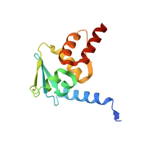
3LBZ

Crystal Structure of the BCL6 BTB domain complexed with the small molecule inhibitor 79-6


Experimental Data Snapshot

  • Method: X-RAY DIFFRACTION
  • Resolution: 2.30 Å
  • R-Value Free: 0.269 
  • R-Value Work: 0.213 
  • R-Value Observed: 0.215 

Starting Model: experimental
View more details

wwPDB Validation   3D Report Full Report


Ligand Structure Quality Assessment 


This is version 1.3 of the entry. See complete history


Literature

A small-molecule inhibitor of BCL6 kills DLBCL cells in vitro and in vivo.

Cerchietti, L.C.Ghetu, A.F.Zhu, X.Da Silva, G.F.Zhong, S.Matthews, M.Bunting, K.L.Polo, J.M.Fares, C.Arrowsmith, C.H.Yang, S.N.Garcia, M.Coop, A.Mackerell, A.D.Prive, G.G.Melnick, A.

(2010) Cancer Cell 17: 400-411

  • DOI: https://doi.org/10.1016/j.ccr.2009.12.050
  • Primary Citation of Related Structures:  
    3LBZ

  • PubMed Abstract: 

    The BCL6 transcriptional repressor is the most frequently involved oncogene in diffuse large B cell lymphoma (DLBCL). We combined computer-aided drug design with functional assays to identify low-molecular-weight compounds that bind to the corepressor binding groove of the BCL6 BTB domain. One such compound disrupted BCL6/corepressor complexes in vitro and in vivo, and was observed by X-ray crystallography and NMR to bind the critical site within the BTB groove. This compound could induce expression of BCL6 target genes and kill BCL6-positive DLBCL cell lines. In xenotransplantation experiments, the compound was nontoxic and potently suppressed DLBCL tumors in vivo. The compound also killed primary DLBCLs from human patients.


  • Organizational Affiliation

    Division of Hematology and Medical Oncology, Department of Medicine, Weill Cornell Medical College, Cornell University, New York, NY 10065, USA.


Macromolecules
Find similar proteins by:  (by identity cutoff)  |  3D Structure
Entity ID: 1
MoleculeChains Sequence LengthOrganismDetailsImage
B-cell lymphoma 6 protein
A, B
127Homo sapiensMutation(s): 3 
Gene Names: BCL5BCL6LAZ3ZBTB27ZNF51
UniProt & NIH Common Fund Data Resources
Find proteins for P41182 (Homo sapiens)
Explore P41182 
Go to UniProtKB:  P41182
PHAROS:  P41182
GTEx:  ENSG00000113916 
Entity Groups  
Sequence Clusters30% Identity50% Identity70% Identity90% Identity95% Identity100% Identity
UniProt GroupP41182
Sequence Annotations
Expand
  • Reference Sequence
Experimental Data & Validation

Experimental Data

  • Method: X-RAY DIFFRACTION
  • Resolution: 2.30 Å
  • R-Value Free: 0.269 
  • R-Value Work: 0.213 
  • R-Value Observed: 0.215 
  • Space Group: C 1 2 1
Unit Cell:
Length ( Å )Angle ( ˚ )
a = 101.712α = 90
b = 38.611β = 114.7
c = 78.817γ = 90
Software Package:
Software NamePurpose
HKL-2000data collection
PHASERphasing
REFMACrefinement
DENZOdata reduction
SCALEPACKdata scaling

Structure Validation

View Full Validation Report


Ligand Structure Quality Assessment 


Entry History 

Deposition Data

Revision History  (Full details and data files)

  • Version 1.0: 2010-04-28
    Type: Initial release
  • Version 1.1: 2011-07-13
    Changes: Version format compliance
  • Version 1.2: 2021-10-13
    Changes: Database references, Derived calculations
  • Version 1.3: 2023-09-06
    Changes: Data collection, Refinement description