


The Crystal Structure of smu.793 from Streptococcus mutans UA159 bound to acetyl CoA
Su, X.-D., Hou, Q.M., Fan, X.X., Nan, J., Liu, X.To be published.
Experimental Data Snapshot
Starting Model: experimental
View more details
Entity ID: 1 | |||||
|---|---|---|---|---|---|
| Molecule | Chains | Sequence Length | Organism | Details | Image |
| Putative uncharacterized protein smu.793 | 163 | Streptococcus mutans UA159 | Mutation(s): 0 Gene Names: smu.793 | 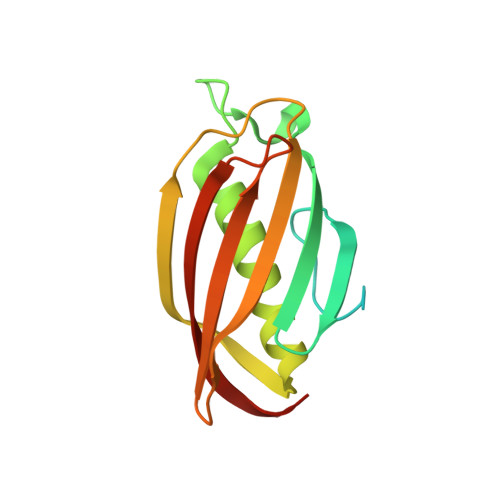 | |
UniProt | |||||
Entity Groups | |||||
| Sequence Clusters | 30% Identity50% Identity70% Identity90% Identity95% Identity100% Identity | ||||
| UniProt Group | Q8DUV0 | ||||
Sequence AnnotationsExpand | |||||
Reference Sequence | |||||
| Ligands 2 Unique | |||||
|---|---|---|---|---|---|
| ID | Chains | Name / Formula / InChI Key | 2D Diagram | 3D Interactions | |
| COA Download:Ideal Coordinates CCD File | E [auth A], G [auth B], J [auth C], K [auth D] | COENZYME A C21 H36 N7 O16 P3 S RGJOEKWQDUBAIZ-IBOSZNHHSA-N |  | ||
| CL Download:Ideal Coordinates CCD File | F [auth A], H [auth B], I [auth B], L [auth D] | CHLORIDE ION Cl VEXZGXHMUGYJMC-UHFFFAOYSA-M |  | ||
| Length ( Å ) | Angle ( ˚ ) |
|---|---|
| a = 68.536 | α = 90 |
| b = 68.536 | β = 90 |
| c = 118.817 | γ = 90 |
| Software Name | Purpose |
|---|---|
| CrystalClear | data collection |
| PHENIX | refinement |


