3L48 | pdb_00003l48

Crystal structure of the C-terminal domain of the PapC usher


Experimental Data Snapshot

  • Method: X-RAY DIFFRACTION
  • Resolution: 2.10 Å
  • R-Value Free: 
    0.230 (Depositor), 0.209 (DCC) 
  • R-Value Work: 
    0.200 (Depositor) 
  • R-Value Observed: 
    0.201 (Depositor) 

wwPDB Validation 3D Report Full Report

Validation slider image for 3L48

This is version 1.2 of the entry. See complete history

Literature

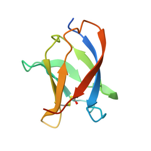
Structural Homology between the C-Terminal Domain of the PapC Usher and Its Plug.

Ford, B.Rego, A.T.Ragan, T.J.Pinkner, J.Dodson, K.Driscoll, P.C.Hultgren, S.Waksman, G.

(2010) J Bacteriol 192: 1824-1831

  • DOI: https://doi.org/10.1128/JB.01677-09
  • Primary Citation Related Structures: 
    2KT6, 3L48

  • PubMed Abstract: 

    P pili are extracellular appendages responsible for the targeting of uropathogenic Escherichia coli to the kidney. They are assembled by the chaperone-usher (CU) pathway of pilus biogenesis involving two proteins, the periplasmic chaperone PapD and the outer membrane assembly platform, PapC. Many aspects of the structural biology of the Pap CU pathway have been elucidated, except for the C-terminal domain of the PapC usher, the structure of which is unknown. In this report, we identify a stable and folded fragment of the C-terminal region of the PapC usher and determine its structure using both X-ray crystallography and nuclear magnetic resonance (NMR) spectroscopy. These structures reveal a beta-sandwich fold very similar to that of the plug domain, a domain of PapC obstructing its translocation domain. This structural similarity suggests similar functions in usher-mediated pilus biogenesis, playing out at different stages of the process. This structure paves the way for further functional analysis targeting surfaces common to both the plug and the C-terminal domain of PapC.


  • Organizational Affiliation
    • Department of Pathology and Immunology, Washington University School of Medicine, Saint Louis, Missouri 63110, USA.

Macromolecule Content 

  • Total Structure Weight: 51.44 kDa 
  • Atom Count: 3,343 
  • Modeled Residue Count: 406 
  • Deposited Residue Count: 470 
  • Unique protein chains: 1

Macromolecules

Find similar proteins by:|  3D Structure
Entity ID: 1
MoleculeChains  Sequence LengthOrganismDetailsImage
Outer membrane usher protein PapC
A, B, C, D, E
94Escherichia coli UTI89Mutation(s): 0 
Gene Names: papCUTI89_C4893
UniProt
Find proteins for Q1R2W8 (Escherichia coli (strain UTI89 / UPEC))
Explore Q1R2W8 
Go to UniProtKB:  Q1R2W8
Entity Groups
Sequence Clusters30% Identity50% Identity70% Identity90% Identity95% Identity100% Identity
UniProt GroupQ1R2W8
Sequence Annotations
Expand
Reference Sequence

Experimental Data & Validation

Experimental Data

  • Method: X-RAY DIFFRACTION
  • Resolution: 2.10 Å
  • R-Value Free:  0.230 (Depositor), 0.209 (DCC) 
  • R-Value Work:  0.200 (Depositor) 
  • R-Value Observed: 0.201 (Depositor) 
Space Group: P 42 21 2
Unit Cell:
Length ( Å )Angle ( ˚ )
a = 100.936α = 90
b = 100.936β = 90
c = 89.54γ = 90
Software Package:
Software NamePurpose
Blu-Icedata collection
PHASERphasing
PHENIXrefinement
XDSdata reduction
XSCALEdata scaling

Structure Validation

View Full Validation Report


Entry History 

Deposition Data

Revision History  (Full details and data files)

  • Version 1.0: 2010-03-02
    Type: Initial release
  • Version 1.1: 2011-07-13
    Changes: Version format compliance
  • Version 1.2: 2024-11-06
    Changes: Data collection, Database references, Derived calculations, Structure summary