3L2I | pdb_00003l2i

1.85 Angstrom Crystal Structure of the 3-Dehydroquinate Dehydratase (aroD) from Salmonella typhimurium LT2.


Experimental Data Snapshot

  • Method: X-RAY DIFFRACTION
  • Resolution: 1.85 Å
  • R-Value Free: 
    0.214 (Depositor), 0.235 (DCC) 
  • R-Value Work: 
    0.168 (Depositor) 
  • R-Value Observed: 
    0.170 (Depositor) 

Starting Model: experimental
View more details

wwPDB Validation 3D Report Full Report

Validation slider image for 3L2I

This is version 1.3 of the entry. See complete history

Literature

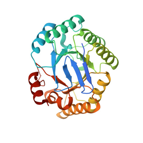
A conserved surface loop in type I dehydroquinate dehydratases positions an active site arginine and functions in substrate binding.

Light, S.H.Minasov, G.Shuvalova, L.Peterson, S.N.Caffrey, M.Anderson, W.F.Lavie, A.

(2011) Biochemistry 50: 2357-2363

  • DOI: https://doi.org/10.1021/bi102020s
  • Primary Citation Related Structures: 
    3L2I, 3O1N, 3OEX

  • PubMed Abstract: 

    Dehydroquinate dehydratase (DHQD) catalyzes the third step in the biosynthetic shikimate pathway. We present three crystal structures of the Salmonella enterica type I DHQD that address the functionality of a surface loop that is observed to close over the active site following substrate binding. Two wild-type structures with differing loop conformations and kinetic and structural studies of a mutant provide evidence of both direct and indirect mechanisms of involvement of the loop in substrate binding. In addition to allowing amino acid side chains to establish a direct interaction with the substrate, closure of the loop necessitates a conformational change of a key active site arginine, which in turn positions the substrate productively. The absence of DHQD in humans and its essentiality in many pathogenic bacteria make the enzyme a target for the development of nontoxic antimicrobials. The structures and ligand binding insights presented here may inform the design of novel type I DHQD inhibiting molecules.


  • Organizational Affiliation
    • Center for Structural Genomics of Infectious Diseases, Feinberg School of Medicine, Northwestern University, Chicago, Illinois 60611, United States.

Macromolecule Content 

  • Total Structure Weight: 60.23 kDa 
  • Atom Count: 4,572 
  • Modeled Residue Count: 509 
  • Deposited Residue Count: 552 
  • Unique protein chains: 1

Macromolecules

Find similar proteins by:|  3D Structure
Entity ID: 1
MoleculeChains  Sequence LengthOrganismDetailsImage
3-dehydroquinate dehydratase
A, B
276Salmonella enterica subsp. enterica serovar Typhimurium str. LT2Mutation(s): 0 
Gene Names: aroDSTM1358
EC: 4.2.1.10
UniProt
Find proteins for P58687 (Salmonella typhimurium (strain LT2 / SGSC1412 / ATCC 700720))
Explore P58687 
Go to UniProtKB:  P58687
Entity Groups
Sequence Clusters30% Identity50% Identity70% Identity90% Identity95% Identity100% Identity
UniProt GroupP58687
Sequence Annotations
Expand
Reference Sequence

Small Molecules

Ligands 1 Unique
IDChains Name / Formula / InChI Key2D Diagram3D Interactions
MG

Query on MG


Download:Ideal Coordinates CCD File
C [auth B]MAGNESIUM ION
Mg
JLVVSXFLKOJNIY-UHFFFAOYSA-N

Experimental Data & Validation

Experimental Data

  • Method: X-RAY DIFFRACTION
  • Resolution: 1.85 Å
  • R-Value Free:  0.214 (Depositor), 0.235 (DCC) 
  • R-Value Work:  0.168 (Depositor) 
  • R-Value Observed: 0.170 (Depositor) 
Space Group: P 21 21 21
Unit Cell:
Length ( Å )Angle ( ˚ )
a = 36.919α = 90
b = 76.909β = 90
c = 171.854γ = 90
Software Package:
Software NamePurpose
Blu-Icedata collection
PHASERphasing
REFMACrefinement
HKL-2000data reduction
HKL-2000data scaling

Structure Validation

View Full Validation Report


Entry History 

Revision History  (Full details and data files)

  • Version 1.0: 2009-12-29
    Type: Initial release
  • Version 1.1: 2011-07-13
    Changes: Version format compliance
  • Version 1.2: 2017-11-01
    Changes: Refinement description
  • Version 1.3: 2023-09-06
    Changes: Data collection, Database references, Derived calculations, Refinement description