3L11 | pdb_00003l11

Crystal Structure of the Ring Domain of RNF168


Experimental Data Snapshot

  • Method: X-RAY DIFFRACTION
  • Resolution: 2.12 Å
  • R-Value Free: 
    0.215 (Depositor), 0.218 (DCC) 
  • R-Value Work: 
    0.187 (Depositor), 0.195 (DCC) 
  • R-Value Observed: 
    0.189 (Depositor) 

Starting Model: experimental
View more details

wwPDB Validation 3D Report Full Report

Validation slider image for 3L11

This is version 1.3 of the entry. See complete history

Literature

Molecular insights into the function of RING finger (RNF)-containing proteins hRNF8 and hRNF168 in Ubc13/Mms2-dependent ubiquitylation.

Campbell, S.J.Edwards, R.A.Leung, C.C.Neculai, D.Hodge, C.D.Dhe-Paganon, S.Glover, J.N.

(2012) J Biological Chem 287: 23900-23910

  • DOI: https://doi.org/10.1074/jbc.M112.359653
  • Primary Citation Related Structures: 
    3L11, 4ORH

  • PubMed Abstract: 

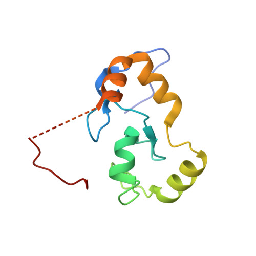
    The repair of DNA double strand breaks by homologous recombination relies on the unique topology of the chains formed by Lys-63 ubiquitylation of chromatin to recruit repair factors such as breast cancer 1 (BRCA1) to sites of DNA damage. The human RING finger (RNF) E3 ubiquitin ligases, RNF8 and RNF168, with the E2 ubiquitin-conjugating complex Ubc13/Mms2, perform the majority of Lys-63 ubiquitylation in homologous recombination. Here, we show that RNF8 dimerizes and binds to Ubc13/Mms2, thereby stimulating formation of Lys-63 ubiquitin chains, whereas the related RNF168 RING domain is a monomer and does not catalyze Lys-63 polyubiquitylation. The crystal structure of the RNF8/Ubc13/Mms2 ternary complex reveals the structural basis for the interaction between Ubc13 and the RNF8 RING and that an extended RNF8 coiled-coil is responsible for its dimerization. Mutations that disrupt the RNF8/Ubc13 binding surfaces, or that truncate the RNF8 coiled-coil, reduce RNF8-catalyzed ubiquitylation. These findings support the hypothesis that RNF8 is responsible for the initiation of Lys-63-linked ubiquitylation in the DNA damage response, which is subsequently amplified by RNF168.


  • Organizational Affiliation
    • Department of Biochemistry, University of Alberta, Edmonton, Alberta, Canada T6G 2H7.

Macromolecule Content 

  • Total Structure Weight: 13.47 kDa 
  • Atom Count: 891 
  • Modeled Residue Count: 104 
  • Deposited Residue Count: 115 
  • Unique protein chains: 1

Macromolecules

Find similar proteins by:|  3D Structure
Entity ID: 1
MoleculeChains  Sequence LengthOrganismDetailsImage
E3 ubiquitin-protein ligase RNF168115Homo sapiensMutation(s): 0 
Gene Names: BC033791RNF168
EC: 6.3.2 (PDB Primary Data), 2.3.2.27 (UniProt)
UniProt & NIH Common Fund Data Resources
Find proteins for Q8IYW5 (Homo sapiens)
Explore Q8IYW5 
Go to UniProtKB:  Q8IYW5
PHAROS:  Q8IYW5
GTEx:  ENSG00000163961 
Entity Groups
Sequence Clusters30% Identity50% Identity70% Identity90% Identity95% Identity100% Identity
UniProt GroupQ8IYW5
Sequence Annotations
Expand
Reference Sequence

Experimental Data & Validation

Experimental Data

  • Method: X-RAY DIFFRACTION
  • Resolution: 2.12 Å
  • R-Value Free:  0.215 (Depositor), 0.218 (DCC) 
  • R-Value Work:  0.187 (Depositor), 0.195 (DCC) 
  • R-Value Observed: 0.189 (Depositor) 
Space Group: P 43 21 2
Unit Cell:
Length ( Å )Angle ( ˚ )
a = 49.706α = 90
b = 49.706β = 90
c = 110.149γ = 90
Software Package:
Software NamePurpose
CrystalCleardata collection
SHELXDphasing
MOLREPphasing
REFMACrefinement
XDSdata reduction
XDSdata scaling

Structure Validation

View Full Validation Report


Entry History 

Revision History  (Full details and data files)

  • Version 1.0: 2010-01-19
    Type: Initial release
  • Version 1.1: 2011-07-13
    Changes: Version format compliance
  • Version 1.2: 2012-09-26
    Changes: Database references
  • Version 1.3: 2023-09-06
    Changes: Data collection, Database references, Derived calculations, Refinement description