3L0W | pdb_00003l0w

Structure of split monoubiquitinated PCNA with ubiquitin in position two


Experimental Data Snapshot

  • Method: X-RAY DIFFRACTION
  • Resolution: 2.80 Å
  • R-Value Free: 
    0.314 (Depositor), 0.325 (DCC) 
  • R-Value Work: 
    0.277 (Depositor), 0.294 (DCC) 
  • R-Value Observed: 
    0.279 (Depositor) 

Starting Model: experimental
View more details

wwPDB Validation 3D Report Full Report

Validation slider image for 3L0W

This is version 1.3 of the entry. See complete history

Literature

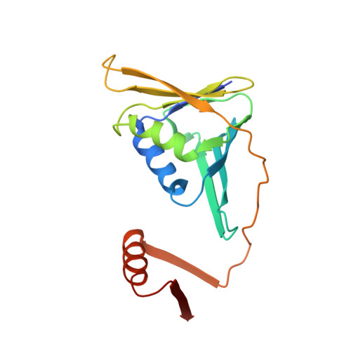
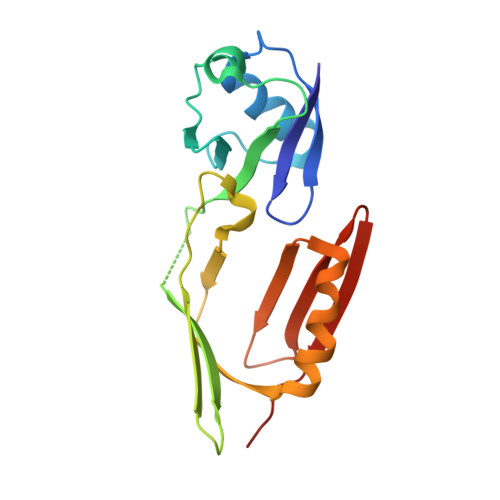
Structure of monoubiquitinated PCNA and implications for translesion synthesis and DNA polymerase exchange.

Freudenthal, B.D.Gakhar, L.Ramaswamy, S.Washington, M.T.

(2010) Nat Struct Mol Biol 17: 479-484

  • DOI: https://doi.org/10.1038/nsmb.1776
  • Primary Citation Related Structures: 
    3L0W, 3L0X, 3L10

  • PubMed Abstract: 

    DNA synthesis by classical polymerases can be blocked by many lesions. These blocks are overcome by translesion synthesis, whereby the stalled classical, replicative polymerase is replaced by a nonclassical polymerase. In eukaryotes this polymerase exchange requires proliferating cell nuclear antigen (PCNA) monoubiquitination. To better understand the polymerase exchange, we developed a means of producing monoubiquitinated PCNA, by splitting the protein into two self-assembling polypeptides. We determined the X-ray crystal structure of monoubiquitinated PCNA and found that the ubiquitin moieties are located on the back face of PCNA and interact with it through their canonical hydrophobic surface. Moreover, the attachment of ubiquitin does not change PCNA's conformation. We propose that PCNA ubiquitination facilitates nonclassical polymerase recruitment to the back of PCNA by forming a new binding surface for nonclassical polymerases, consistent with a 'tool belt' model of the polymerase exchange.


  • Organizational Affiliation
    • Department of Biochemistry, University of Iowa College of Medicine, Iowa City, Iowa, USA.

Macromolecule Content 

  • Total Structure Weight: 37.95 kDa 
  • Atom Count: 2,590 
  • Modeled Residue Count: 329 
  • Deposited Residue Count: 338 
  • Unique protein chains: 2

Macromolecules

Find similar proteins by:|  3D Structure
Entity ID: 1
MoleculeChains  Sequence LengthOrganismDetailsImage
Proliferating cell nuclear antigen169Saccharomyces cerevisiaeMutation(s): 0 
Gene Names: POL30YBR0811YBR088C
UniProt
Find proteins for P15873 (Saccharomyces cerevisiae (strain ATCC 204508 / S288c))
Explore P15873 
Go to UniProtKB:  P15873
Entity Groups
Sequence Clusters30% Identity50% Identity70% Identity90% Identity95% Identity100% Identity
UniProt GroupP15873
Sequence Annotations
Expand
Reference Sequence
Find similar proteins by:|  3D Structure
Entity ID: 2
MoleculeChains  Sequence LengthOrganismDetailsImage
Monoubiquitinated Proliferating cell nuclear antigen169Saccharomyces cerevisiaeMutation(s): 0 
Gene Names: POL30UBI1 & Pol30YBR0811YBR088C
UniProt
Find proteins for P05759 (Saccharomyces cerevisiae (strain ATCC 204508 / S288c))
Explore P05759 
Go to UniProtKB:  P05759
Find proteins for P15873 (Saccharomyces cerevisiae (strain ATCC 204508 / S288c))
Explore P15873 
Go to UniProtKB:  P15873
Entity Groups
Sequence Clusters30% Identity50% Identity70% Identity90% Identity95% Identity100% Identity
UniProt GroupsP15873P05759
Sequence Annotations
Expand
Reference Sequence

Experimental Data & Validation

Experimental Data

  • Method: X-RAY DIFFRACTION
  • Resolution: 2.80 Å
  • R-Value Free:  0.314 (Depositor), 0.325 (DCC) 
  • R-Value Work:  0.277 (Depositor), 0.294 (DCC) 
  • R-Value Observed: 0.279 (Depositor) 
Space Group: P 21 3
Unit Cell:
Length ( Å )Angle ( ˚ )
a = 122.516α = 90
b = 122.516β = 90
c = 122.516γ = 90
Software Package:
Software NamePurpose
PHASERphasing
REFMACrefinement
PDB_EXTRACTdata extraction
StructureStudiodata collection
d*TREKdata reduction
d*TREKdata scaling

Structure Validation

View Full Validation Report


Entry History 

Deposition Data

Revision History  (Full details and data files)

  • Version 1.0: 2010-03-23
    Type: Initial release
  • Version 1.1: 2011-07-13
    Changes: Version format compliance
  • Version 1.2: 2017-08-02
    Changes: Refinement description, Source and taxonomy
  • Version 1.3: 2023-09-06
    Changes: Data collection, Database references, Refinement description