3KW0 | pdb_00003kw0

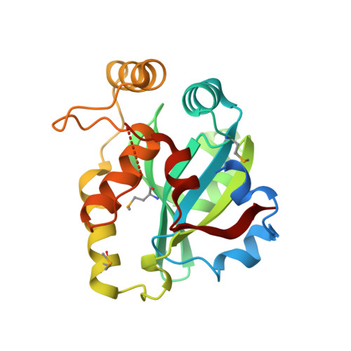
Crystal structure of Cysteine peptidase (NP_982244.1) from BACILLUS CEREUS ATCC 10987 at 2.50 A resolution


Experimental Data Snapshot

  • Method: X-RAY DIFFRACTION
  • Resolution: 2.50 Å
  • R-Value Free: 
    0.219 (Depositor), 0.218 (DCC) 
  • R-Value Work: 
    0.192 (Depositor), 0.195 (DCC) 
  • R-Value Observed: 
    0.193 (Depositor) 

wwPDB Validation 3D Report Full Report

Validation slider image for 3KW0

This is version 1.6 of the entry. See complete history. 

Literature

Structural Analysis of Papain-Like NlpC/P60 Superfamily Enzymes with a Circularly Permuted Topology Reveals Potential Lipid Binding Sites.

Xu, Q., Rawlings, N.D., Chiu, H.J., Jaroszewski, L., Klock, H.E., Knuth, M.W., Miller, M.D., Elsliger, M.A., Deacon, A.M., Godzik, A., Lesley, S.A., Wilson, I.A.

(2011) PLoS One 6: e22013-e22013

  • DOI: https://doi.org/10.1371/journal.pone.0022013
  • Primary Citation Related Structures: 
    3KW0

  • PubMed Abstract: 

    NlpC/P60 superfamily papain-like enzymes play important roles in all kingdoms of life. Two members of this superfamily, LRAT-like and YaeF/YiiX-like families, were predicted to contain a catalytic domain that is circularly permuted such that the catalytic cysteine is located near the C-terminus, instead of at the N-terminus. These permuted enzymes are widespread in virus, pathogenic bacteria, and eukaryotes. We determined the crystal structure of a member of the YaeF/YiiX-like family from Bacillus cereus in complex with lysine. The structure, which adopts a ligand-induced, "closed" conformation, confirms the circular permutation of catalytic residues. A comparative analysis of other related protein structures within the NlpC/P60 superfamily is presented. Permutated NlpC/P60 enzymes contain a similar conserved core and arrangement of catalytic residues, including a Cys/His-containing triad and an additional conserved tyrosine. More surprisingly, permuted enzymes have a hydrophobic S1 binding pocket that is distinct from previously characterized enzymes in the family, indicative of novel substrate specificity. Further analysis of a structural homolog, YiiX (PDB 2if6) identified a fatty acid in the conserved hydrophobic pocket, thus providing additional insights into possible function of these novel enzymes.


  • Organizational Affiliation: 
    • Joint Center for Structural Genomics, La Jolla, California, United States of America.

Macromolecule Content 

  • Total Structure Weight: 100.16 kDa 
  • Atom Count: 6,102 
  • Modeled Residue Count: 773 
  • Deposited Residue Count: 856 
  • Unique protein chains: 1

Macromolecules

Find similar proteins by:|  3D Structure
Entity ID: 1
MoleculeChains  Sequence LengthOrganismDetailsImage
Cysteine peptidase
A, B, C, D
214Bacillus cereus ATCC 10987Mutation(s): 0 
Gene Names: BCE_A0238
UniProt
Find proteins for Q74NK7 (Bacillus cereus (strain ATCC 10987 / NRS 248))
Explore Q74NK7 
Go to UniProtKB:  Q74NK7
Entity Groups
Sequence Clusters30% Identity50% Identity70% Identity90% Identity95% Identity100% Identity
UniProt GroupQ74NK7
Sequence Annotations
Expand
Reference Sequence

Small Molecules

Ligands 2 Unique
IDChains Name / Formula / InChI Key2D Diagram3D Interactions
LYS

Query on LYS


Download:Ideal Coordinates CCD File
E [auth A],
I [auth B],
M [auth C],
P [auth D]
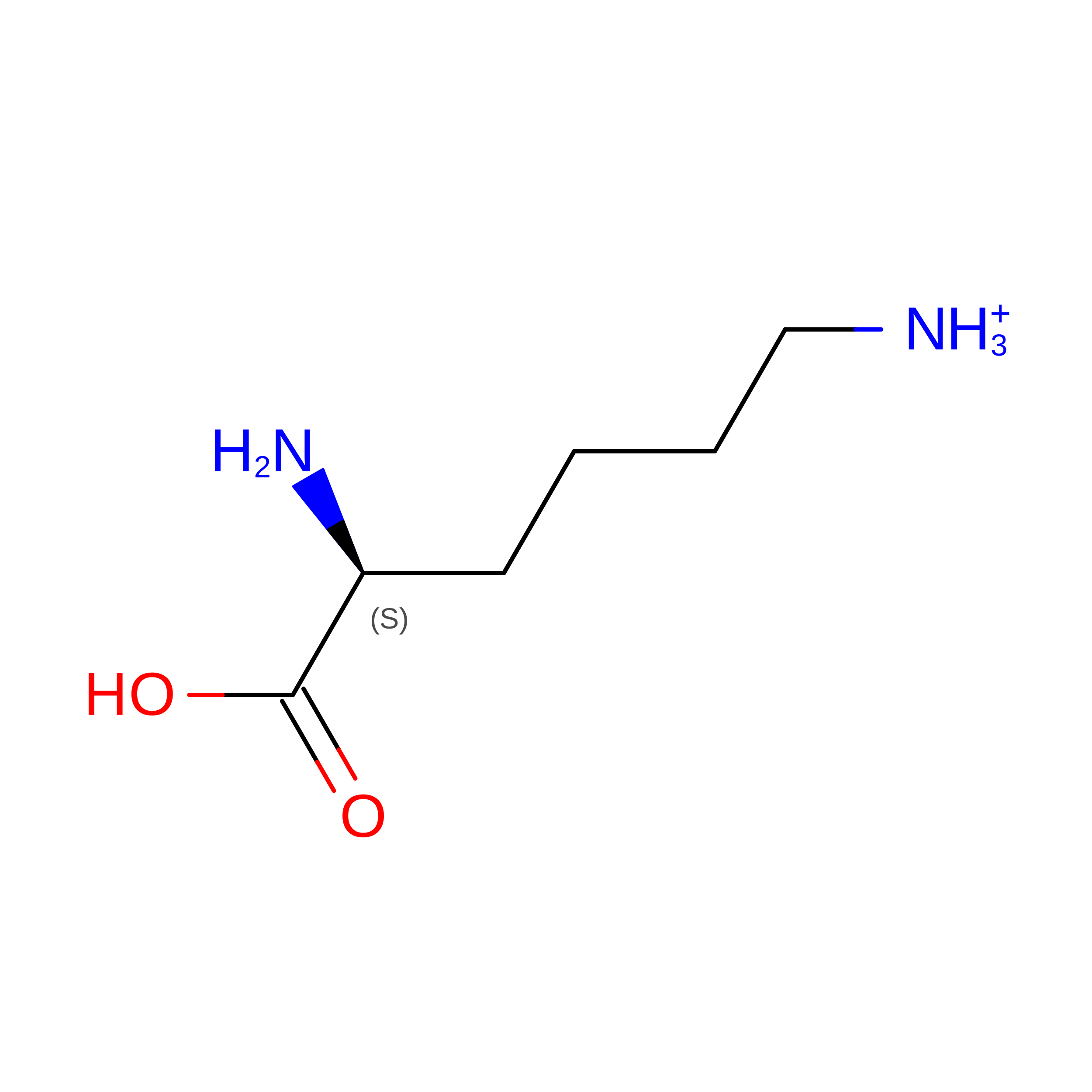
LYSINE
C6 H15 N2 O2
KDXKERNSBIXSRK-YFKPBYRVSA-O
CL

Query on CL


Download:Ideal Coordinates CCD File
F [auth A]
G [auth A]
H [auth A]
J [auth B]
K [auth B]
F [auth A],
G [auth A],
H [auth A],
J [auth B],
K [auth B],
L [auth B],
N [auth C],
O [auth C],
Q [auth D]
CHLORIDE ION
Cl
VEXZGXHMUGYJMC-UHFFFAOYSA-M
Modified Residues  1 Unique
IDChains TypeFormula2D DiagramParent
MSE
Query on MSE
A, B, C, D
L-PEPTIDE LINKINGC5 H11 N O2 SeMET

Experimental Data & Validation

Experimental Data

  • Method: X-RAY DIFFRACTION
  • Resolution: 2.50 Å
  • R-Value Free:  0.219 (Depositor), 0.218 (DCC) 
  • R-Value Work:  0.192 (Depositor), 0.195 (DCC) 
  • R-Value Observed: 0.193 (Depositor) 
Space Group: P 65
Unit Cell:
Length ( Å )Angle ( ˚ )
a = 64.96α = 90
b = 64.96β = 90
c = 407.8γ = 120
Software Package:
Software NamePurpose
REFMACrefinement
PHENIXrefinement
SHELXphasing
MolProbitymodel building
XSCALEdata scaling
PDB_EXTRACTdata extraction
XDSdata reduction
SHELXDphasing
autoSHARPphasing

Structure Validation

View Full Validation Report


Entry History 

Deposition Data

Revision History  (Full details and data files)

  • Version 1.0: 2009-12-15
    Type: Initial release
  • Version 1.1: 2011-07-13
    Changes: Advisory, Source and taxonomy, Version format compliance
  • Version 1.2: 2011-08-10
    Changes: Database references
  • Version 1.3: 2017-11-01
    Changes: Refinement description
  • Version 1.4: 2019-07-17
    Changes: Data collection, Derived calculations, Refinement description
  • Version 1.5: 2023-02-01
    Changes: Database references, Derived calculations
  • Version 1.6: 2024-11-20
    Changes: Data collection, Refinement description, Structure summary