3KPF | pdb_00003kpf

X-ray structure of the mutant Lys300Met of polyamine oxidase from Zea mays


Experimental Data Snapshot

  • Method: X-RAY DIFFRACTION
  • Resolution: 2.90 Å
  • R-Value Free: 
    0.223 (Depositor), 0.213 (DCC) 
  • R-Value Work: 
    0.186 (Depositor), 0.178 (DCC) 
  • R-Value Observed: 
    0.188 (Depositor) 

Starting Model: experimental
View more details

wwPDB Validation 3D Report Full Report

Validation slider image for 3KPF

Ligand Structure Quality Assessment 


This is version 2.3 of the entry. See complete history

Literature

The crystal structure of the mutant K300M of polyamine oxidase from ZEA MAYS unveils the role of LYS300 in catalysis

Fiorillo, A.Ilari, A.Tavladoraki, P.

To be published.

Macromolecule Content 

  • Total Structure Weight: 111.53 kDa 
  • Atom Count: 7,861 
  • Modeled Residue Count: 931 
  • Deposited Residue Count: 956 
  • Unique protein chains: 1

Macromolecules

Find similar proteins by:|  3D Structure
Entity ID: 1
MoleculeChains  Sequence LengthOrganismDetailsImage
Polyamine oxidase
A, B
478Zea maysMutation(s): 1 
Gene Names: PAO
EC: 1.5.3 (PDB Primary Data), 1.5.3.14 (UniProt), 1.5.3.15 (UniProt)
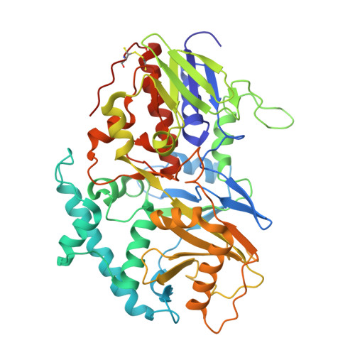
UniProt
Find proteins for O64411 (Zea mays)
Explore O64411 
Go to UniProtKB:  O64411
Entity Groups
Sequence Clusters30% Identity50% Identity70% Identity90% Identity95% Identity100% Identity
UniProt GroupO64411
Glycosylation
Glycosylation Sites: 1
Sequence Annotations
Expand
Reference Sequence

Oligosaccharides

Help  
Entity ID: 2
MoleculeChains Length2D Diagram GlycosylationD Interactions
2-acetamido-2-deoxy-beta-D-glucopyranose-(1-4)-2-acetamido-2-deoxy-beta-D-glucopyranose
C
2N-Glycosylation
Glycosylation Resources
GlyTouCan: G42666HT
GlyCosmos: G42666HT
GlyGen: G42666HT

Small Molecules

Ligands 5 Unique
IDChains Name / Formula / InChI Key2D Diagram3D Interactions
FAD

Query on FAD


Download:Ideal Coordinates CCD File
D [auth A],
H [auth B]
FLAVIN-ADENINE DINUCLEOTIDE
C27 H33 N9 O15 P2
VWWQXMAJTJZDQX-UYBVJOGSSA-N
NAG

Query on NAG


Download:Ideal Coordinates CCD File
E [auth A]2-acetamido-2-deoxy-beta-D-glucopyranose
C8 H15 N O6
OVRNDRQMDRJTHS-FMDGEEDCSA-N
SO4

Query on SO4


Download:Ideal Coordinates CCD File
I [auth B]SULFATE ION
O4 S
QAOWNCQODCNURD-UHFFFAOYSA-L
ACT

Query on ACT


Download:Ideal Coordinates CCD File
G [auth A],
J [auth B]
ACETATE ION
C2 H3 O2
QTBSBXVTEAMEQO-UHFFFAOYSA-M
CL

Query on CL


Download:Ideal Coordinates CCD File
F [auth A]CHLORIDE ION
Cl
VEXZGXHMUGYJMC-UHFFFAOYSA-M

Experimental Data & Validation

Experimental Data

  • Method: X-RAY DIFFRACTION
  • Resolution: 2.90 Å
  • R-Value Free:  0.223 (Depositor), 0.213 (DCC) 
  • R-Value Work:  0.186 (Depositor), 0.178 (DCC) 
  • R-Value Observed: 0.188 (Depositor) 
Space Group: P 31 2 1
Unit Cell:
Length ( Å )Angle ( ˚ )
a = 138.606α = 90
b = 138.606β = 90
c = 189.471γ = 120
Software Package:
Software NamePurpose
MOLREPphasing
REFMACrefinement
DENZOdata reduction
SCALEPACKdata scaling

Structure Validation

View Full Validation Report


Ligand Structure Quality Assessment 


Entry History 

Deposition Data

Revision History  (Full details and data files)

  • Version 1.0: 2010-11-17
    Type: Initial release
  • Version 1.1: 2011-07-13
    Changes: Version format compliance
  • Version 2.0: 2020-07-29
    Type: Remediation
    Reason: Carbohydrate remediation
    Changes: Advisory, Atomic model, Data collection, Database references, Derived calculations, Structure summary
  • Version 2.1: 2021-10-13
    Changes: Advisory, Database references, Structure summary
  • Version 2.2: 2023-09-06
    Changes: Data collection, Refinement description
  • Version 2.3: 2024-10-30
    Changes: Structure summary