3KP3 | pdb_00003kp3

Staphylococcus epidermidis in complex with ampicillin


Experimental Data Snapshot

  • Method: X-RAY DIFFRACTION
  • Resolution: 3.20 Å
  • R-Value Free: 
    0.274 (Depositor), 0.270 (DCC) 
  • R-Value Work: 
    0.247 (Depositor), 0.234 (DCC) 

Starting Model: experimental
View more details

wwPDB Validation 3D Report Full Report

Validation slider image for 3KP3

Ligand Structure Quality Assessment 


This is version 1.2 of the entry. See complete history

Literature

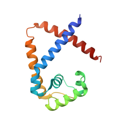
Structural study of TcaR and its complexes with multiple antibiotics from Staphylococcus epidermidis.

Chang, Y.M.Jeng, W.Y.Ko, T.P.Yeh, Y.J.Chen, C.K.Wang, A.H.

(2010) Proc Natl Acad Sci U S A 107: 8617-8622

  • DOI: https://doi.org/10.1073/pnas.0913302107
  • Primary Citation Related Structures: 
    3KP2, 3KP3, 3KP4, 3KP5, 3KP6, 3KP7

  • PubMed Abstract: 

    TcaR and IcaR are a weak and a strong negative regulator of transcription of the ica locus, respectively, and their presence prevents the poly-N-acetylglucosamine production and biofilm formation in Staphylococcus epidermidis. Although TcaR was shown to interact with the ica promoter, the precise binding region and the mechanism of interaction remained unclear. Here we present the 3D structure of TcaR in its apo form and in complex with salicylate as well as several aminoglycoside and beta-lactam antibiotics. A comparison of the native and complex TcaR structures indicates that the mechanism of regulation involves a large conformational change in the DNA-binding lobe. Here, we deduced the consensus binding sequence of two [ approximately TTNNAA] hexamers embedded in a 16 bp sequence for a TcaR dimer. Six TcaR dimers bind specifically to three approximately 33 bp segments close to the IcaR binding region with varying affinities, and their repressor activity is directly interfered by salicylate and different classes of natural antimicrobial compounds. We also found in this study that the antimicrobial compounds we tested were shown not only to inhibit TcaR-DNA interaction but also to further induce biofilm formation in S. epidermidis in our in vivo assay. The results support a general mechanism for antibiotics in regulating TcaR-DNA interaction and thereby help understand the effect of antibiotic exposure on bacterial antibiotic resistance through biofilm formation.


  • Organizational Affiliation
    • Institute of Biochemical Sciences, National Taiwan University, Taipei 106, Taiwan.

Macromolecule Content 

  • Total Structure Weight: 35.46 kDa 
  • Atom Count: 2,322 
  • Modeled Residue Count: 273 
  • Deposited Residue Count: 302 
  • Unique protein chains: 1

Macromolecules

Find similar proteins by:|  3D Structure
Entity ID: 1
MoleculeChains  Sequence LengthOrganismDetailsImage
Transcriptional regulator TcaR
A, B
151Staphylococcus epidermidis RP62AMutation(s): 0 
Gene Names: SERP1949SE_1937tcaR
UniProt
Find proteins for Q5HLN6 (Staphylococcus epidermidis (strain ATCC 35984 / DSM 28319 / BCRC 17069 / CCUG 31568 / BM 3577 / RP62A))
Explore Q5HLN6 
Go to UniProtKB:  Q5HLN6
Entity Groups
Sequence Clusters30% Identity50% Identity70% Identity90% Identity95% Identity100% Identity
UniProt GroupQ5HLN6
Sequence Annotations
Expand
Reference Sequence

Small Molecules

Ligands 1 Unique
IDChains Name / Formula / InChI Key2D Diagram3D Interactions
AIC

Query on AIC


Download:Ideal Coordinates CCD File
C [auth B],
D [auth B]
(2S,5R,6R)-6-{[(2R)-2-AMINO-2-PHENYLETHANOYL]AMINO}-3,3-DIMETHYL-7-OXO-4-THIA-1-AZABICYCLO[3.2.0]HEPTANE-2-CARBOXYLIC ACID
C16 H19 N3 O4 S
AVKUERGKIZMTKX-NJBDSQKTSA-N

Experimental Data & Validation

Experimental Data

  • Method: X-RAY DIFFRACTION
  • Resolution: 3.20 Å
  • R-Value Free:  0.274 (Depositor), 0.270 (DCC) 
  • R-Value Work:  0.247 (Depositor), 0.234 (DCC) 
Space Group: P 61
Unit Cell:
Length ( Å )Angle ( ˚ )
a = 108.033α = 90
b = 108.033β = 90
c = 49.76γ = 120
Software Package:
Software NamePurpose
HKL-2000data collection
CNSrefinement
HKL-2000data reduction
HKL-2000data scaling
CNSphasing

Structure Validation

View Full Validation Report


Ligand Structure Quality Assessment 


Entry History 

Deposition Data

Revision History  (Full details and data files)

  • Version 1.0: 2010-06-09
    Type: Initial release
  • Version 1.1: 2011-07-13
    Changes: Version format compliance
  • Version 1.2: 2023-11-01
    Changes: Data collection, Database references, Derived calculations, Refinement description