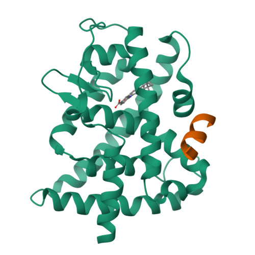
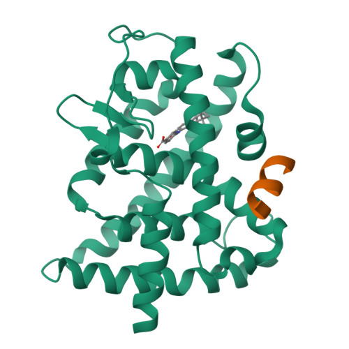
3KMR

Crystal structure of RARalpha ligand binding domain in complex with an agonist ligand (Am580) and a coactivator fragment


Experimental Data Snapshot

  • Method: X-RAY DIFFRACTION
  • Resolution: 1.80 Å
  • R-Value Free: 0.237 
  • R-Value Work: 0.197 
  • R-Value Observed: 0.199 

Starting Model: experimental
View more details

wwPDB Validation   3D Report Full Report


Ligand Structure Quality Assessment 


This is version 1.2 of the entry. See complete history


Literature

A unique secondary-structure switch controls constitutive gene repression by retinoic acid receptor.

le Maire, A.Teyssier, C.Erb, C.Grimaldi, M.Alvarez, S.de Lera, A.R.Balaguer, P.Gronemeyer, H.Royer, C.A.Germain, P.Bourguet, W.

(2010) Nat Struct Mol Biol 17: 801-807

  • DOI: https://doi.org/10.1038/nsmb.1855
  • Primary Citation of Related Structures:  
    3KMR, 3KMZ

  • PubMed Abstract: 

    In the absence of ligand, some nuclear receptors, including retinoic acid receptor (RAR), act as transcriptional repressors by recruiting corepressor complexes to target genes. This constitutive repression is crucial in metazoan reproduction, development and homeostasis. However, its specific molecular determinants had remained obscure. Using structural, biochemical and cell-based assays, we show that the basal repressive activity of RAR is conferred by an extended beta-strand that forms an antiparallel beta-sheet with specific corepressor residues. Agonist binding induces a beta-strand-to-alpha-helix transition that allows for helix H11 formation, which in turn provokes corepressor release, repositioning of helix H12 and coactivator recruitment. Several lines of evidence suggest that this structural switch could be implicated in the intrinsic repressor function of other nuclear receptors. Finally, we report on the molecular mechanism by which inverse agonists strengthen corepressor interaction and enhance gene silencing by RAR.


  • Organizational Affiliation

    Institut National de la Santé et de la Recherche Médicale, U554, Montpellier, France.


Macromolecules
Find similar proteins by:  (by identity cutoff)  |  3D Structure
Entity ID: 1
MoleculeChains Sequence LengthOrganismDetailsImage
Retinoic acid receptor alpha266Homo sapiensMutation(s): 0 
Gene Names: NR1B1RARA
UniProt & NIH Common Fund Data Resources
Find proteins for P10276 (Homo sapiens)
Explore P10276 
Go to UniProtKB:  P10276
PHAROS:  P10276
GTEx:  ENSG00000131759 
Entity Groups  
Sequence Clusters30% Identity50% Identity70% Identity90% Identity95% Identity100% Identity
UniProt GroupP10276
Sequence Annotations
Expand
  • Reference Sequence

Find similar proteins by:  Sequence   |   3D Structure  

Entity ID: 2
MoleculeChains Sequence LengthOrganismDetailsImage
Nuclear receptor coactivator 1B [auth C]13N/AMutation(s): 0 
EC: 2.3.1.48
UniProt & NIH Common Fund Data Resources
Find proteins for Q15788 (Homo sapiens)
Explore Q15788 
Go to UniProtKB:  Q15788
PHAROS:  Q15788
GTEx:  ENSG00000084676 
Entity Groups  
Sequence Clusters30% Identity50% Identity70% Identity90% Identity95% Identity100% Identity
UniProt GroupQ15788
Sequence Annotations
Expand
  • Reference Sequence
Small Molecules
Ligands 1 Unique
IDChains Name / Formula / InChI Key2D Diagram3D Interactions
EQN
Query on EQN

Download Ideal Coordinates CCD File 
C [auth A]4-{[(5,5,8,8-tetramethyl-5,6,7,8-tetrahydronaphthalen-2-yl)carbonyl]amino}benzoic acid
C22 H25 N O3
SZWKGOZKRMMLAJ-UHFFFAOYSA-N
Binding Affinity Annotations 
IDSourceBinding Affinity
EQN BindingDB:  3KMR Kd: min: 36, max: 1400 (nM) from 3 assay(s)
IC50: 41 (nM) from 1 assay(s)
EC50: min: 0.3, max: 79.43 (nM) from 10 assay(s)
Experimental Data & Validation

Experimental Data

  • Method: X-RAY DIFFRACTION
  • Resolution: 1.80 Å
  • R-Value Free: 0.237 
  • R-Value Work: 0.197 
  • R-Value Observed: 0.199 
  • Space Group: C 1 2 1
Unit Cell:
Length ( Å )Angle ( ˚ )
a = 88.307α = 90
b = 61.218β = 105.38
c = 49.444γ = 90
Software Package:
Software NamePurpose
ADSCdata collection
MOLREPphasing
REFMACrefinement
MOSFLMdata reduction
SCALAdata scaling

Structure Validation

View Full Validation Report


Ligand Structure Quality Assessment 


Entry History 

Deposition Data

Revision History  (Full details and data files)

  • Version 1.0: 2010-06-02
    Type: Initial release
  • Version 1.1: 2011-07-13
    Changes: Version format compliance
  • Version 1.2: 2023-09-06
    Changes: Data collection, Database references, Derived calculations, Refinement description