3KLZ | pdb_00003klz

Pentameric formate channel with formate bound


Experimental Data Snapshot

  • Method: X-RAY DIFFRACTION
  • Resolution: 2.50 Å
  • R-Value Free: 
    0.223 (Depositor), 0.215 (DCC) 
  • R-Value Work: 
    0.173 (Depositor), 0.173 (DCC) 
  • R-Value Observed: 
    0.175 (Depositor) 

wwPDB Validation 3D Report Full Report

Validation slider image for 3KLZ

Ligand Structure Quality Assessment 


This is version 1.3 of the entry. See complete history

Literature

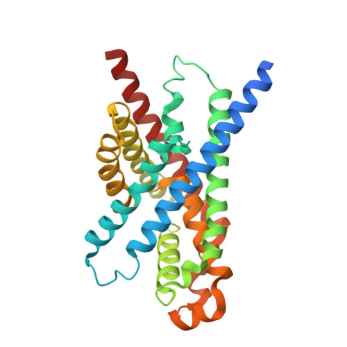
Structure and mechanism of a pentameric formate channel

Waight, A.B.Love, J.Wang, D.N.

(2010) Nat Struct Mol Biol 17: 31-37

  • DOI: https://doi.org/10.1038/nsmb.1740
  • Primary Citation Related Structures: 
    3KLY, 3KLZ

  • PubMed Abstract: 

    Formate transport across the inner membrane is a critical step in anaerobic bacterial respiration. Members of the formate/nitrite transport protein family function to shuttle substrate across the cytoplasmic membrane. In bacterial pathogens, the nitrite transport protein is involved in protecting bacteria from peroxynitrite released by host macrophages. We have determined the 2.13-A structure of the formate channel FocA from Vibrio cholerae, which reveals a pentamer in which each monomer possesses its own substrate translocation pore. Unexpectedly, the fold of the FocA monomer resembles that found in water and glycerol channels. The selectivity filter in FocA consists of a cytoplasmic slit and a central constriction ring. A 2.5-A high-formate structure shows two formate ions bound to the cytoplasmic slit via both hydrogen bonding and van der Waals interactions, providing a structural basis for the substrate selectivity of the channel.


  • Organizational Affiliation
    • The Helen L. and Martin S. Kimmel Center for Biology and Medicine, Skirball Institute of Biomolecular Medicine, New York University School of Medicine, New York, New York, USA.

Macromolecule Content 

  • Total Structure Weight: 158.77 kDa 
  • Atom Count: 10,446 
  • Modeled Residue Count: 1,281 
  • Deposited Residue Count: 1,400 
  • Unique protein chains: 1

Macromolecules

Find similar proteins by:|  3D Structure
Entity ID: 1
MoleculeChains  Sequence LengthOrganismDetailsImage
Putative formate transporter 1
A, B, C, D, E
280Vibrio choleraeMutation(s): 0 
Gene Names: VC_1695
Membrane Entity: Yes 
UniProt
Find proteins for Q9KRE7 (Vibrio cholerae serotype O1 (strain ATCC 39315 / El Tor Inaba N16961))
Explore Q9KRE7 
Go to UniProtKB:  Q9KRE7
Entity Groups
Sequence Clusters30% Identity50% Identity70% Identity90% Identity95% Identity100% Identity
UniProt GroupQ9KRE7
Sequence Annotations
Expand
Reference Sequence

Small Molecules

Ligands 2 Unique
IDChains Name / Formula / InChI Key2D Diagram3D Interactions
BOG

Query on BOG


Download:Ideal Coordinates CCD File
F [auth A]
G [auth A]
GA [auth C]
H [auth A]
HA [auth C]
F [auth A],
G [auth A],
GA [auth C],
H [auth A],
HA [auth C],
OA [auth D],
R [auth B],
S [auth B],
T [auth B],
U [auth B],
V [auth B],
W [auth B],
WA [auth E],
ZA [auth E]
octyl beta-D-glucopyranoside
C14 H28 O6
HEGSGKPQLMEBJL-RKQHYHRCSA-N
FMT

Query on FMT


Download:Ideal Coordinates CCD File
AA [auth B]
AB [auth E]
BA [auth B]
BB [auth E]
CA [auth B]
AA [auth B],
AB [auth E],
BA [auth B],
BB [auth E],
CA [auth B],
CB [auth E],
DA [auth B],
DB [auth E],
EA [auth B],
EB [auth E],
FA [auth B],
FB [auth E],
I [auth A],
IA [auth C],
J [auth A],
JA [auth C],
K [auth A],
KA [auth C],
L [auth A],
LA [auth C],
M [auth A],
MA [auth C],
N [auth A],
NA [auth C],
O [auth A],
P [auth A],
PA [auth D],
Q [auth A],
QA [auth D],
RA [auth D],
SA [auth D],
TA [auth D],
UA [auth D],
VA [auth D],
X [auth B],
XA [auth E],
Y [auth B],
YA [auth E],
Z [auth B]
FORMIC ACID
C H2 O2
BDAGIHXWWSANSR-UHFFFAOYSA-N

Experimental Data & Validation

Experimental Data

  • Method: X-RAY DIFFRACTION
  • Resolution: 2.50 Å
  • R-Value Free:  0.223 (Depositor), 0.215 (DCC) 
  • R-Value Work:  0.173 (Depositor), 0.173 (DCC) 
  • R-Value Observed: 0.175 (Depositor) 
Space Group: P 21 21 21
Unit Cell:
Length ( Å )Angle ( ˚ )
a = 99.779α = 90
b = 100.456β = 90
c = 192.955γ = 90
Software Package:
Software NamePurpose
PHENIXrefinement
HKL-2000data reduction
SCALEPACKdata scaling
PHENIXphasing

Structure Validation

View Full Validation Report


Ligand Structure Quality Assessment 


Entry History 

Deposition Data

Revision History  (Full details and data files)

  • Version 1.0: 2009-12-15
    Type: Initial release
  • Version 1.1: 2011-07-13
    Changes: Version format compliance
  • Version 1.2: 2020-07-29
    Type: Remediation
    Reason: Carbohydrate remediation
    Changes: Advisory, Data collection, Derived calculations, Structure summary
  • Version 1.3: 2024-02-21
    Changes: Data collection, Database references, Structure summary