3KKR | pdb_00003kkr

Crystal structure of catalytic core domain of BIV integrase in crystal form I


Experimental Data Snapshot

  • Method: X-RAY DIFFRACTION
  • Resolution: 2.45 Å
  • R-Value Free: 
    0.242 (Depositor), 0.244 (DCC) 
  • R-Value Work: 
    0.192 (Depositor), 0.194 (DCC) 
  • R-Value Observed: 
    0.194 (Depositor) 

Starting Model: experimental
View more details

wwPDB Validation 3D Report Full Report

Validation slider image for 3KKR

This is version 1.3 of the entry. See complete history

Literature

Crystal structures of catalytic core domain of BIV integrase: implications for the interaction between integrase and target DNA

Yao, X.Fang, S.Qiao, W.Geng, Y.Shen, Y.

(2010) Protein Cell 1: 363-370

  • DOI: https://doi.org/10.1007/s13238-010-0047-5
  • Primary Citation Related Structures: 
    3KKR, 3KKS

  • PubMed Abstract: 

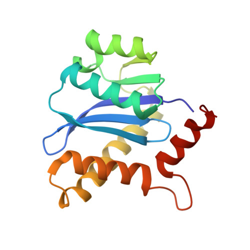
    Integrase plays a critical role in the recombination of viral DNA into the host genome. Therefore, over the past decade, it has been a hot target of drug design in the fight against type 1 human immunodeficiency virus (HIV-1). Bovine immunodeficiency virus (BIV) integrase has the same function as HIV-1 integrase. We have determined crystal structures of the BIV integrase catalytic core domain (CCD) in two different crystal forms at a resolution of 2.45 Å and 2.2 Å, respectively. In crystal form I, BIV integrase CCD forms a back-to-back dimer, in which the two active sites are on opposite sides. This has also been seen in many of the CCD structures of HIV-1 integrase that were determined previously. However, in crystal form II, BIV integrase CCD forms a novel face-to-face dimer in which the two active sites are close to each other. Strikingly, the distance separating the two active sites is approximately 20 Å, a distance that perfectly matches a 5-base pair interval. Based on these data, we propose a model for the interaction of integrase with its target DNA, which is also supported by many published biochemical data. Our results provide important clues for designing new inhibitors against HIV-1.


  • Organizational Affiliation
    • Tianjin Key Laboratory of Protein Science, College of Life Sciences, Nankai University, 94 Weijin Road, Tianjin, 300071, China.

Macromolecule Content 

  • Total Structure Weight: 17.38 kDa 
  • Atom Count: 1,333 
  • Modeled Residue Count: 152 
  • Deposited Residue Count: 152 
  • Unique protein chains: 1

Macromolecules

Find similar proteins by:|  3D Structure
Entity ID: 1
MoleculeChains  Sequence LengthOrganismDetailsImage
Integrase152Bovine immunodeficiency virus R29Mutation(s): 0 
Gene Names: gag-pol
UniProt
Find proteins for P19560 (Bovine immunodeficiency virus (strain R29))
Explore P19560 
Go to UniProtKB:  P19560
Entity Groups
Sequence Clusters30% Identity50% Identity70% Identity90% Identity95% Identity100% Identity
UniProt GroupP19560
Sequence Annotations
Expand
Reference Sequence

Experimental Data & Validation

Experimental Data

  • Method: X-RAY DIFFRACTION
  • Resolution: 2.45 Å
  • R-Value Free:  0.242 (Depositor), 0.244 (DCC) 
  • R-Value Work:  0.192 (Depositor), 0.194 (DCC) 
  • R-Value Observed: 0.194 (Depositor) 
Space Group: P 41 21 2
Unit Cell:
Length ( Å )Angle ( ˚ )
a = 82.32α = 90
b = 82.32β = 90
c = 51.057γ = 90
Software Package:
Software NamePurpose
HKL-2000data collection
PHASERphasing
PHENIXrefinement
HKL-2000data reduction
HKL-2000data scaling

Structure Validation

View Full Validation Report


Entry History 

Deposition Data

  • Released Date: 2010-09-22 
  • Deposition Author(s): Shen, Y.

Revision History  (Full details and data files)

  • Version 1.0: 2010-09-22
    Type: Initial release
  • Version 1.1: 2011-07-13
    Changes: Source and taxonomy, Version format compliance
  • Version 1.2: 2012-03-28
    Changes: Database references
  • Version 1.3: 2023-11-01
    Changes: Data collection, Database references, Derived calculations, Refinement description