3KIN | pdb_00003kin

KINESIN (DIMERIC) FROM RATTUS NORVEGICUS


Experimental Data Snapshot

  • Method: X-RAY DIFFRACTION
  • Resolution: 3.10 Å
  • R-Value Free: 
    0.362 (Depositor) 
  • R-Value Observed: 
    0.289 (Depositor) 

wwPDB Validation 3D Report Full Report

Validation slider image for 3KIN

This is version 1.3 of the entry. See complete history

Literature

The crystal structure of dimeric kinesin and implications for microtubule-dependent motility.

Kozielski, F.Sack, S.Marx, A.Thormahlen, M.Schonbrunn, E.Biou, V.Thompson, A.Mandelkow, E.M.Mandelkow, E.

(1997) Cell 91: 985-994

  • DOI: https://doi.org/10.1016/s0092-8674(00)80489-4
  • Primary Citation Related Structures: 
    3KIN

  • PubMed Abstract: 

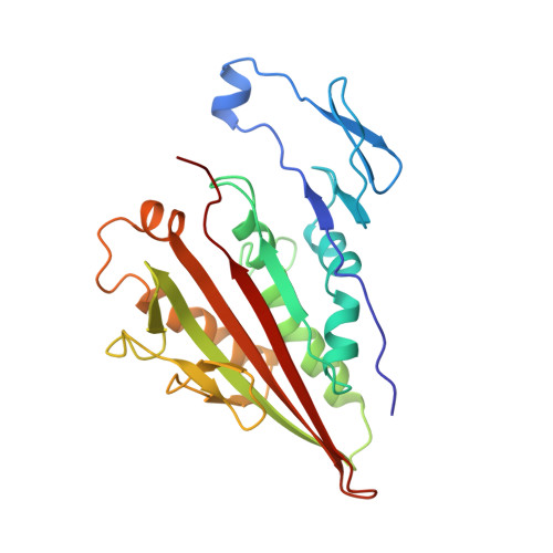
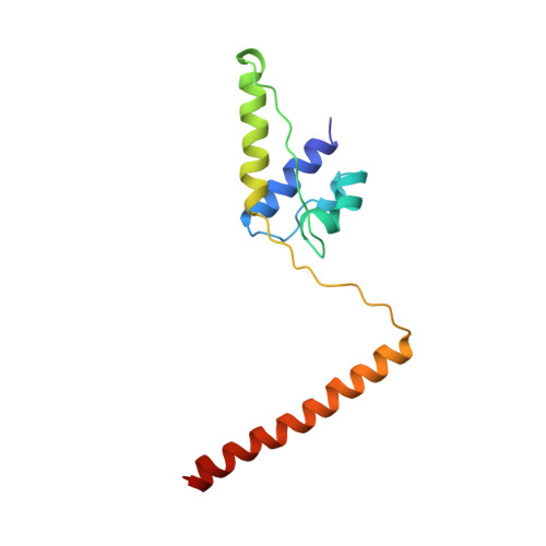
    The dimeric form of the kinesin motor and neck domain from rat brain with bound ADP has been solved by X-ray crystallography. The two heads of the dimer are connected via a coiled-coil alpha-helical interaction of their necks. They are broadly similar to one another; differences are most apparent in the head-neck junction and in a moderate reorientation of the neck helices in order to adopt to the coiled-coil conformation. The heads show a rotational symmetry (approximately 120 degrees) about an axis close to that of the coiled-coil. This arrangement is unexpected since it is not compatible with the microtubule lattice. In this arrangement, the two heads of a kinesin dimer could not have equivalent interactions with microtubules.


  • Organizational Affiliation
    • Max-Planck-Unit for Structural Molecular Biology, Hamburg, Germany.

Macromolecule Content 

  • Total Structure Weight: 81.5 kDa 
  • Atom Count: 5,743 
  • Modeled Residue Count: 708 
  • Deposited Residue Count: 710 
  • Unique protein chains: 2

Macromolecules

Find similar proteins by:|  3D Structure
Entity ID: 1
MoleculeChains  Sequence LengthOrganismDetailsImage
KINESIN HEAVY CHAIN
A, C
238Rattus norvegicusMutation(s): 0 
EC: 3.6.4
UniProt
Find proteins for P56536 (Rattus norvegicus)
Explore P56536 
Go to UniProtKB:  P56536
Entity Groups
Sequence Clusters30% Identity50% Identity70% Identity90% Identity95% Identity100% Identity
UniProt GroupP56536
Sequence Annotations
Expand
Reference Sequence
Find similar proteins by:|  3D Structure
Entity ID: 2
MoleculeChains  Sequence LengthOrganismDetailsImage
KINESIN HEAVY CHAIN
B, D
117Rattus norvegicusMutation(s): 0 
EC: 5.6.1.3
UniProt
Find proteins for Q6QLM7 (Rattus norvegicus)
Explore Q6QLM7 
Go to UniProtKB:  Q6QLM7
Entity Groups
Sequence Clusters30% Identity50% Identity70% Identity90% Identity95% Identity100% Identity
UniProt GroupQ6QLM7
Sequence Annotations
Expand
Reference Sequence

Small Molecules

Ligands 1 Unique
IDChains Name / Formula / InChI Key2D Diagram3D Interactions
ADP

Query on ADP


Download:Ideal Coordinates CCD File
E [auth A],
F [auth C]
ADENOSINE-5'-DIPHOSPHATE
C10 H15 N5 O10 P2
XTWYTFMLZFPYCI-KQYNXXCUSA-N

Experimental Data & Validation

Experimental Data

  • Method: X-RAY DIFFRACTION
  • Resolution: 3.10 Å
  • R-Value Free:  0.362 (Depositor) 
  • R-Value Observed: 0.289 (Depositor) 
Space Group: P 21 21 21
Unit Cell:
Length ( Å )Angle ( ˚ )
a = 72.15α = 90
b = 91.85β = 90
c = 141.68γ = 90
Software Package:
Software NamePurpose
PHASESphasing
REFMACrefinement
DENZOdata reduction
SCALEPACKdata scaling

Structure Validation

View Full Validation Report


Entry History 

Deposition Data

Revision History  (Full details and data files)

  • Version 1.0: 1998-10-14
    Type: Initial release
  • Version 1.1: 2008-03-25
    Changes: Version format compliance
  • Version 1.2: 2011-07-13
    Changes: Version format compliance
  • Version 1.3: 2024-12-25
    Changes: Advisory, Data collection, Database references, Derived calculations, Structure summary