3KHQ | pdb_00003khq

Crystal structure of murine Ig-beta (CD79b) in the monomeric form


Experimental Data Snapshot

  • Method: X-RAY DIFFRACTION
  • Resolution: 1.70 Å
  • R-Value Free: 
    0.197 (Depositor), 0.205 (DCC) 
  • R-Value Work: 
    0.187 (Depositor), 0.197 (DCC) 
  • R-Value Observed: 
    0.188 (Depositor) 

wwPDB Validation 3D Report Full Report

Validation slider image for 3KHQ

Ligand Structure Quality Assessment 


This is version 1.4 of the entry. See complete history

Literature

Structural and Functional Studies of Igalphabeta and Its Assembly with the B Cell Antigen Receptor.

Radaev, S.Zou, Z.Tolar, P.Nguyen, K.Nguyen, A.Krueger, P.D.Stutzman, N.Pierce, S.Sun, P.D.

(2010) Structure 18: 934-943

  • DOI: https://doi.org/10.1016/j.str.2010.04.019
  • Primary Citation Related Structures: 
    3KG5, 3KHO, 3KHQ

  • PubMed Abstract: 

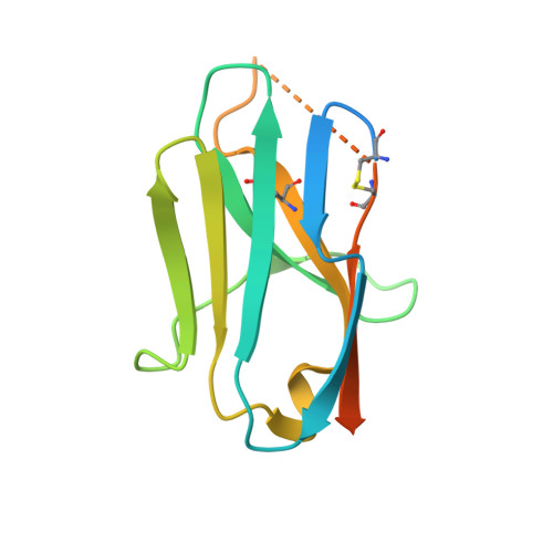
    The B cell antigen receptor (BCR) plays an essential role in all phases of B cell development. Here we show that the extracellular domains of murine and human Igbeta form an I-set immunoglobulin-like structure with an interchain disulfide between cysteines on their G strands. Structural and sequence analysis suggests that Igalpha displays a similar fold as Igbeta. An Igalphabeta heterodimer model was generated based on the unique disulfide-bonded Igbeta dimer. Solution binding studies showed that the extracellular domains of Igalphabeta preferentially recognize the constant region of BCR with mu chain specificity, suggesting a role for Igalphabeta to enhance BCRmu chain signaling. Cluster mutations on Igalpha, Igbeta, and a membrane-bound form of immunoglobulin (mIgM) based on the structural model identified distinct areas of potential contacts involving charged residues on both subunits of the coreceptor and the Cmu4 domain of mIgM. These studies provide the first structural model for understanding BCR function.


  • Organizational Affiliation
    • Structural Immunology Section, Laboratory of Immunogenetics, National Institute of Allergy and Infectious Diseases, National Institutes of Health, 12441 Parklawn Drive, Rockville, MD 20852, USA.

Macromolecule Content 

  • Total Structure Weight: 15.6 kDa 
  • Atom Count: 849 
  • Modeled Residue Count: 93 
  • Deposited Residue Count: 133 
  • Unique protein chains: 1

Macromolecules

Find similar proteins by:|  3D Structure
Entity ID: 1
MoleculeChains  Sequence LengthOrganismDetailsImage
B-cell antigen receptor complex-associated protein beta chain133Mus musculusMutation(s): 0 
Gene Names: Cd79bIgb
UniProt
Find proteins for P15530 (Mus musculus)
Explore P15530 
Go to UniProtKB:  P15530
Entity Groups
Sequence Clusters30% Identity50% Identity70% Identity90% Identity95% Identity100% Identity
UniProt GroupP15530
Sequence Annotations
Expand
Reference Sequence

Biologically Interesting Molecules (External Reference) 

1 Unique

Experimental Data & Validation

Experimental Data

  • Method: X-RAY DIFFRACTION
  • Resolution: 1.70 Å
  • R-Value Free:  0.197 (Depositor), 0.205 (DCC) 
  • R-Value Work:  0.187 (Depositor), 0.197 (DCC) 
  • R-Value Observed: 0.188 (Depositor) 
Space Group: P 21 21 2
Unit Cell:
Length ( Å )Angle ( ˚ )
a = 35.63α = 90
b = 79.72β = 90
c = 34.32γ = 90
Software Package:
Software NamePurpose
REFMACrefinement
CNSrefinement
HKL-2000data reduction
HKL-2000data scaling
CNSphasing

Structure Validation

View Full Validation Report


Ligand Structure Quality Assessment 


Entry History 

Deposition Data

Revision History  (Full details and data files)

  • Version 1.0: 2010-08-25
    Type: Initial release
  • Version 1.1: 2011-07-13
    Changes: Version format compliance
  • Version 1.2: 2011-12-14
    Changes: Non-polymer description
  • Version 1.3: 2019-07-17
    Changes: Advisory, Data collection, Refinement description
  • Version 1.4: 2025-03-26
    Changes: Advisory, Data collection, Database references, Derived calculations, Structure summary