3KDS | pdb_00003kds

apo-FtsH crystal structure


Experimental Data Snapshot

  • Method: X-RAY DIFFRACTION
  • Resolution: 2.60 Å
  • R-Value Free: 
    0.287 (Depositor), 0.289 (DCC) 
  • R-Value Work: 
    0.223 (Depositor), 0.224 (DCC) 
  • R-Value Observed: 
    0.226 (Depositor) 

Starting Model: experimental
View more details

wwPDB Validation 3D Report Full Report

Validation slider image for 3KDS

Ligand Structure Quality Assessment 


This is version 1.4 of the entry. See complete history

Literature

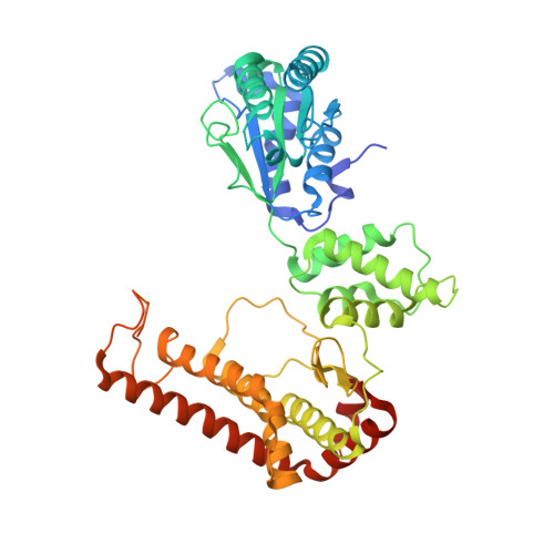
The crystal structure of apo-FtsH reveals domain movements necessary for substrate unfolding and translocation

Bieniossek, C.Niederhauser, B.Baumann, U.M.

(2009) Proc Natl Acad Sci U S A 106: 21579-21584

  • DOI: https://doi.org/10.1073/pnas.0910708106
  • Primary Citation Related Structures: 
    3KDS

  • PubMed Abstract: 

    The hexameric membrane-spanning ATP-dependent metalloprotease FtsH is universally conserved in eubacteria, mitochondria, and chloroplasts, where it fulfills key functions in quality control and signaling. As a member of the self-compartmentalizing ATPases associated with various cellular activities (AAA+ proteases), FtsH converts the chemical energy stored in ATP via conformational rearrangements into a mechanical force that is used for substrate unfolding and translocation into the proteolytic chamber. The crystal structure of the ADP state of Thermotoga maritima FtsH showed a hexameric assembly consisting of a 6-fold symmetric protease disk and a 2-fold symmetric AAA ring. The 2.6 A resolution structure of the cytosolic region of apo-FtsH presented here reveals a new arrangement where the ATPase ring shows perfect 6-fold symmetry with the crucial pore residues lining an open circular entrance. Triggered by this conformational change, a substrate-binding edge beta strand appears within the proteolytic domain. Comparison of the apo- and ADP-bound structure visualizes an inward movement of the aromatic pore residues and generates a model of substrate translocation by AAA+ proteases. Furthermore, we demonstrate that mutation of a conserved glycine in the linker region inactivates FtsH.


  • Organizational Affiliation
    • Department of Chemistry and Biochemistry, University of Bern, Freiestrasse 3, CH-3012 Bern, Switzerland.

Macromolecule Content 

  • Total Structure Weight: 155.81 kDa 
  • Atom Count: 9,994 
  • Modeled Residue Count: 1,279 
  • Deposited Residue Count: 1,395 
  • Unique protein chains: 1

Macromolecules

Find similar proteins by:|  3D Structure
Entity ID: 1
MoleculeChains  Sequence LengthOrganismDetailsImage
Cell division protein FtsHA [auth E],
B [auth F],
C [auth G]
465Thermotoga maritimaMutation(s): 3 
Gene Names: FtsH
EC: 3.4.24
Membrane Entity: Yes 
UniProt
Find proteins for Q9WZ49 (Thermotoga maritima (strain ATCC 43589 / DSM 3109 / JCM 10099 / NBRC 100826 / MSB8))
Explore Q9WZ49 
Go to UniProtKB:  Q9WZ49
Entity Groups
Sequence Clusters30% Identity50% Identity70% Identity90% Identity95% Identity100% Identity
UniProt GroupQ9WZ49
Sequence Annotations
Expand
Reference Sequence

Experimental Data & Validation

Experimental Data

  • Method: X-RAY DIFFRACTION
  • Resolution: 2.60 Å
  • R-Value Free:  0.287 (Depositor), 0.289 (DCC) 
  • R-Value Work:  0.223 (Depositor), 0.224 (DCC) 
  • R-Value Observed: 0.226 (Depositor) 
Space Group: P 6 2 2
Unit Cell:
Length ( Å )Angle ( ˚ )
a = 190.5α = 90
b = 190.5β = 90
c = 152.13γ = 120
Software Package:
Software NamePurpose
XSCALEdata scaling
PHASERphasing
PHENIXrefinement
PDB_EXTRACTdata extraction
DNAdata collection
XDSdata reduction

Structure Validation

View Full Validation Report


Ligand Structure Quality Assessment 


Entry History 

Deposition Data

Revision History  (Full details and data files)

  • Version 1.0: 2009-12-01
    Type: Initial release
  • Version 1.1: 2011-07-13
    Changes: Version format compliance
  • Version 1.2: 2013-01-16
    Changes: Database references
  • Version 1.3: 2021-11-10
    Changes: Database references, Derived calculations
  • Version 1.4: 2023-11-01
    Changes: Data collection, Refinement description