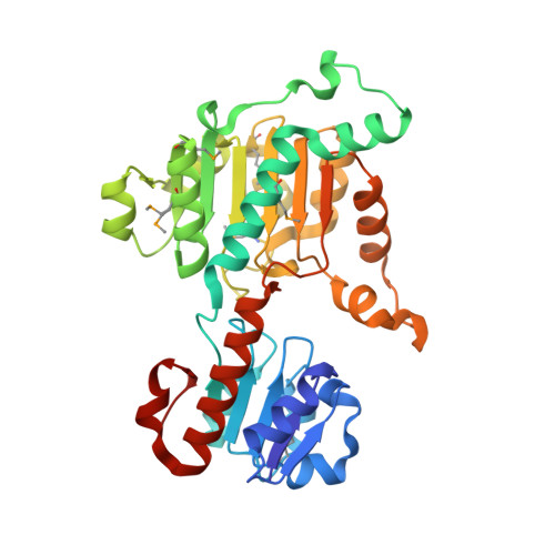
3KB6

Crystal structure of D-Lactate dehydrogenase from aquifex aeolicus complexed with NAD and Lactic acid


Experimental Data Snapshot

  • Method: X-RAY DIFFRACTION
  • Resolution: 2.12 Å
  • R-Value Free: 0.212 
  • R-Value Work: 0.164 
  • R-Value Observed: 0.167 

Starting Model: experimental
View more details

wwPDB Validation   3D Report Full Report


Ligand Structure Quality Assessment 


This is version 2.0 of the entry. See complete history


Literature

Structure of D-lactate dehydrogenase from Aquifex aeolicus complexed with NAD(+) and lactic acid (or pyruvate).

Antonyuk, S.V.Strange, R.W.Ellis, M.J.Bessho, Y.Kuramitsu, S.Inoue, Y.Yokoyama, S.Hasnain, S.S.

(2009) Acta Crystallogr Sect F Struct Biol Cryst Commun 65: 1209-1213

  • DOI: https://doi.org/10.1107/S1744309109044935
  • Primary Citation of Related Structures:  
    3KB6

  • PubMed Abstract: 

    The crystal structure of D-lactate dehydrogenase from Aquifex aeolicus (aq_727) was determined to 2.12 A resolution in space group P2(1)2(1)2(1), with unit-cell parameters a = 90.94, b = 94.43, c = 188.85 A. The structure was solved by molecular replacement using the coenzyme-binding domain of Lactobacillus helveticus D-lactate dehydrogenase and contained two homodimers in the asymmetric unit. Each subunit of the homodimer was found to be in a ;closed' conformation with the NADH cofactor bound to the coenzyme-binding domain and with a lactate (or pyruvate) molecule bound at the interdomain active-site cleft.


  • Organizational Affiliation

    Molecular Biophysics Group, School of Biological Sciences, University of Liverpool, Crown Street, Liverpool L69 7ZB, England.


Macromolecules
Find similar proteins by:  (by identity cutoff)  |  3D Structure
Entity ID: 1
MoleculeChains Sequence LengthOrganismDetailsImage
D-lactate dehydrogenase
A, B, C, D
334Aquifex aeolicusMutation(s): 0 
Gene Names: aq_727ldhA
EC: 1.1.1.28
UniProt
Find proteins for O66939 (Aquifex aeolicus (strain VF5))
Explore O66939 
Go to UniProtKB:  O66939
Entity Groups  
Sequence Clusters30% Identity50% Identity70% Identity90% Identity95% Identity100% Identity
UniProt GroupO66939
Sequence Annotations
Expand
  • Reference Sequence
Small Molecules
Ligands 6 Unique
IDChains Name / Formula / InChI Key2D Diagram3D Interactions
NAD
Query on NAD

Download Ideal Coordinates CCD File 
G [auth A],
JA [auth C],
QA [auth D],
Y [auth B]
NICOTINAMIDE-ADENINE-DINUCLEOTIDE
C21 H27 N7 O14 P2
BAWFJGJZGIEFAR-NNYOXOHSSA-N
1PE
Query on 1PE

Download Ideal Coordinates CCD File 
AA [auth B]
BA [auth B]
FA [auth B]
I [auth A]
L [auth A]
AA [auth B],
BA [auth B],
FA [auth B],
I [auth A],
L [auth A],
LA [auth C],
N [auth A],
Q [auth A]
PENTAETHYLENE GLYCOL
C10 H22 O6
JLFNLZLINWHATN-UHFFFAOYSA-N
PEG
Query on PEG

Download Ideal Coordinates CCD File 
GA [auth C]
H [auth A]
K [auth A]
M [auth A]
NA [auth C]
GA [auth C],
H [auth A],
K [auth A],
M [auth A],
NA [auth C],
R [auth A],
S [auth A],
T [auth A],
TA [auth D],
U [auth A]
DI(HYDROXYETHYL)ETHER
C4 H10 O3
MTHSVFCYNBDYFN-UHFFFAOYSA-N
GOL
Query on GOL

Download Ideal Coordinates CCD File 
CA [auth B]
DA [auth B]
EA [auth B]
KA [auth C]
MA [auth C]
CA [auth B],
DA [auth B],
EA [auth B],
KA [auth C],
MA [auth C],
O [auth A],
P [auth A],
RA [auth D],
SA [auth D],
V [auth B],
Z [auth B]
GLYCEROL
C3 H8 O3
PEDCQBHIVMGVHV-UHFFFAOYSA-N
LAC
Query on LAC

Download Ideal Coordinates CCD File 
F [auth A],
IA [auth C],
PA [auth D],
X [auth B]
LACTIC ACID
C3 H6 O3
JVTAAEKCZFNVCJ-UWTATZPHSA-N
PPI
Query on PPI

Download Ideal Coordinates CCD File 
E [auth A],
HA [auth C],
J [auth A],
OA [auth D],
W [auth B]
PROPANOIC ACID
C3 H6 O2
XBDQKXXYIPTUBI-UHFFFAOYSA-N
Modified Residues  1 Unique
IDChains TypeFormula2D DiagramParent
MSE
Query on MSE
A, B, C, D
L-PEPTIDE LINKINGC5 H11 N O2 SeMET
Experimental Data & Validation

Experimental Data

  • Method: X-RAY DIFFRACTION
  • Resolution: 2.12 Å
  • R-Value Free: 0.212 
  • R-Value Work: 0.164 
  • R-Value Observed: 0.167 
  • Space Group: P 21 21 21
Unit Cell:
Length ( Å )Angle ( ˚ )
a = 90.938α = 90
b = 94.432β = 90
c = 188.849γ = 90
Software Package:
Software NamePurpose
DENZOdata reduction
SCALEPACKdata scaling
MOLREPphasing
REFMACrefinement
PDB_EXTRACTdata extraction
HKL-2000data collection

Structure Validation

View Full Validation Report


Ligand Structure Quality Assessment 


Entry History 

Deposition Data

Revision History  (Full details and data files)

  • Version 1.0: 2009-11-10
    Type: Initial release
  • Version 1.1: 2011-07-13
    Changes: Non-polymer description, Version format compliance
  • Version 1.2: 2017-11-01
    Changes: Advisory, Refinement description
  • Version 1.3: 2023-11-01
    Changes: Advisory, Data collection, Database references, Derived calculations, Refinement description
  • Version 2.0: 2023-11-15
    Changes: Atomic model, Data collection