3K5I | pdb_00003k5i

Crystal structure of N5-carboxyaminoimidazole synthase from aspergillus clavatus in complex with ADP and 5-aminoimadazole ribonucleotide


Experimental Data Snapshot

  • Method: X-RAY DIFFRACTION
  • Resolution: 2.00 Å
  • R-Value Free: 
    0.269 (Depositor), 0.203 (DCC) 
  • R-Value Work: 
    0.203 (Depositor), 0.207 (DCC) 
  • R-Value Observed: 
    0.206 (Depositor) 

Starting Model: experimental
View more details

wwPDB Validation 3D Report Full Report

Validation slider image for 3K5I

Ligand Structure Quality Assessment 


This is version 2.0 of the entry. See complete history

Literature

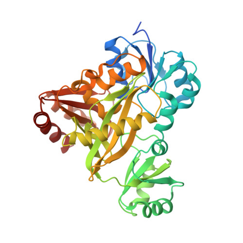
Structural and functional studies of Aspergillus clavatus N(5)-carboxyaminoimidazole ribonucleotide synthetase

Thoden, J.B.Holden, H.M.Paritala, H.Firestine, S.M.

(2010) Biochemistry 49: 752-760

  • DOI: https://doi.org/10.1021/bi901599u
  • Primary Citation Related Structures: 
    3K5H, 3K5I

  • PubMed Abstract: 

    N(5)-Carboxyaminoimidazole ribonucleotide synthetase (N(5)-CAIR synthetase), a key enzyme in microbial de novo purine biosynthesis, catalyzes the conversion of aminoimidazole ribonucleotide (AIR) to N(5)-CAIR. To date, this enzyme has been observed only in microorganisms, and thus, it represents an ideal target for antimicrobial drug development. Here we report the cloning, crystallization, and three-dimensional structural analysis of Aspergillus clavatus N(5)-CAIR synthetase solved in the presence of either Mg(2)ATP or MgADP and AIR. These structures, determined to 2.1 and 2.0 A, respectively, revealed that AIR binds in a pocket analogous to that observed for other ATP-grasp enzymes involved in purine metabolism. On the basis of these models, a site-directed mutagenesis study was subsequently conducted that focused on five amino acid residues located in the active site region of the enzyme. These investigations demonstrated that Asp 153 and Lys 353 play critical roles in catalysis without affecting substrate binding. All other mutations affected substrate binding and, in some instances, catalysis as well. Taken together, the structural and kinetic data presented here suggest a catalytic mechanism whereby Mg(2)ATP and bicarbonate first react to form the unstable intermediate carboxyphosphate. This intermediate subsequently decarboxylates to CO(2) and inorganic phosphate, and the amino group of AIR, through general base assistance by Asp 153, attacks CO(2) to form N(5)-CAIR.


  • Organizational Affiliation
    • Department of Biochemistry, University of Wisconsin, Madison, Wisconsin 53706, USA.

Macromolecule Content 

  • Total Structure Weight: 182.34 kDa 
  • Atom Count: 13,147 
  • Modeled Residue Count: 1,512 
  • Deposited Residue Count: 1,612 
  • Unique protein chains: 1

Macromolecules

Find similar proteins by:|  3D Structure
Entity ID: 1
MoleculeChains  Sequence LengthOrganismDetailsImage
Phosphoribosyl-aminoimidazole carboxylase
A, B, C, D
403Aspergillus clavatusMutation(s): 0 
Gene Names: ACLA_051590ade2
EC: 4.1.1.21
UniProt
Find proteins for A1CII2 (Aspergillus clavatus (strain ATCC 1007 / CBS 513.65 / DSM 816 / NCTC 3887 / NRRL 1 / QM 1276 / 107))
Explore A1CII2 
Go to UniProtKB:  A1CII2
Entity Groups
Sequence Clusters30% Identity50% Identity70% Identity90% Identity95% Identity100% Identity
UniProt GroupA1CII2
Sequence Annotations
Expand
Reference Sequence

Small Molecules

Ligands 5 Unique
IDChains Name / Formula / InChI Key2D Diagram3D Interactions
ADP

Query on ADP


Download:Ideal Coordinates CCD File
G [auth A],
J [auth B],
N [auth C],
Q [auth D]
ADENOSINE-5'-DIPHOSPHATE
C10 H15 N5 O10 P2
XTWYTFMLZFPYCI-KQYNXXCUSA-N
AIR

Query on AIR


Download:Ideal Coordinates CCD File
I [auth A],
L [auth B],
P [auth C],
S [auth D]
5-AMINOIMIDAZOLE RIBONUCLEOTIDE
C8 H14 N3 O7 P
PDACUKOKVHBVHJ-XVFCMESISA-N
NHE

Query on NHE


Download:Ideal Coordinates CCD File
F [auth A],
M [auth C]
2-[N-CYCLOHEXYLAMINO]ETHANE SULFONIC ACID
C8 H17 N O3 S
MKWKNSIESPFAQN-UHFFFAOYSA-N
MG

Query on MG


Download:Ideal Coordinates CCD File
H [auth A],
K [auth B],
O [auth C],
R [auth D]
MAGNESIUM ION
Mg
JLVVSXFLKOJNIY-UHFFFAOYSA-N
NA

Query on NA


Download:Ideal Coordinates CCD File
E [auth A]SODIUM ION
Na
FKNQFGJONOIPTF-UHFFFAOYSA-N

Experimental Data & Validation

Experimental Data

  • Method: X-RAY DIFFRACTION
  • Resolution: 2.00 Å
  • R-Value Free:  0.269 (Depositor), 0.203 (DCC) 
  • R-Value Work:  0.203 (Depositor), 0.207 (DCC) 
  • R-Value Observed: 0.206 (Depositor) 
Space Group: P 1 21 1
Unit Cell:
Length ( Å )Angle ( ˚ )
a = 76.5α = 90
b = 134.2β = 105.6
c = 98.5γ = 90
Software Package:
Software NamePurpose
PROTEUM PLUSdata collection
PHASERphasing
TNTrefinement
SAINTdata reduction
SADABSdata scaling

Structure Validation

View Full Validation Report


Ligand Structure Quality Assessment 


Entry History 

Deposition Data

Revision History  (Full details and data files)

  • Version 1.0: 2009-10-20
    Type: Initial release
  • Version 1.1: 2011-07-13
    Changes: Version format compliance
  • Version 1.2: 2018-03-21
    Changes: Structure summary
  • Version 1.3: 2023-09-06
    Changes: Data collection, Database references, Derived calculations, Refinement description
  • Version 2.0: 2024-01-31
    Changes: Atomic model