3K55 | pdb_00003k55

Structure of beta hairpin deletion mutant of beta toxin from Staphylococcus aureus


Experimental Data Snapshot

  • Method: X-RAY DIFFRACTION
  • Resolution: 3.35 Å
  • R-Value Free: 
    0.281 (Depositor), 0.279 (DCC) 
  • R-Value Work: 
    0.241 (Depositor), 0.241 (DCC) 
  • R-Value Observed: 
    0.243 (Depositor) 

Starting Model: experimental
View more details

wwPDB Validation 3D Report Full Report

Validation slider image for 3K55

This is version 1.3 of the entry. See complete history

Literature

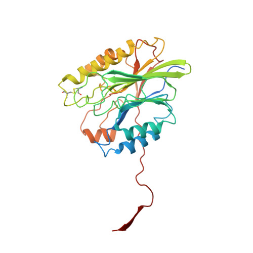
Structure of a mutant beta toxin from Staphylococcus aureus reveals domain swapping and conformational flexibility

Kruse, A.C.Huseby, M.J.Shi, K.Digre, J.Ohlendorf, D.H.Earhart, C.A.

(2011) Acta Crystallogr Sect F Struct Biol Cryst Commun 67: 438-441

  • DOI: https://doi.org/10.1107/S1744309111005239
  • Primary Citation Related Structures: 
    3K55

  • PubMed Abstract: 

    The 3.35 Å resolution crystal structure of a mutant form of the staphylococcal sphingomyelinase β toxin in which a conserved hydrophobic β-hairpin has been deleted is reported. It is shown that this mutation induces domain swapping of a C-terminal β-strand, leading to the formation of dimers linked by a conformationally flexible hinge region. Eight dimers are seen in the asymmetric unit, exhibiting a broad spectrum of conformations trapped in place by intermolecular contacts within the crystal lattice. Furthermore, the 16 monomers within each asymmetric unit exhibit a remarkable heterogeneity in thermal factors, which can be accounted for by the varying degrees to which each monomer interacts with other molecules in the crystal. This structure provides a unique example of the challenges associated with crystallographic study of flexible proteins.


  • Organizational Affiliation
    • Department of Biochemistry, Molecular Biology and Biophysics, University of Minnesota, Minneapolis, MN 55455, USA.

Macromolecule Content 

  • Total Structure Weight: 556.12 kDa 
  • Atom Count: 35,844 
  • Modeled Residue Count: 4,480 
  • Deposited Residue Count: 4,896 
  • Unique protein chains: 1

Macromolecules

Find similar proteins by:|  3D Structure
Entity ID: 1
MoleculeChains  Sequence LengthOrganismDetailsImage
Beta-hemolysin
A, B, C, D, E
A, B, C, D, E, F, G, H, I, J, K, L, M, N, O, P
306Staphylococcus aureus subsp. aureus RN4220Mutation(s): 0 
Gene Names: hlb
EC: 3.1.4.12
UniProt
Find proteins for A7LAI8 (Staphylococcus aureus)
Explore A7LAI8 
Go to UniProtKB:  A7LAI8
Entity Groups
Sequence Clusters30% Identity50% Identity70% Identity90% Identity95% Identity100% Identity
UniProt GroupA7LAI8
Sequence Annotations
Expand
Reference Sequence

Experimental Data & Validation

Experimental Data

  • Method: X-RAY DIFFRACTION
  • Resolution: 3.35 Å
  • R-Value Free:  0.281 (Depositor), 0.279 (DCC) 
  • R-Value Work:  0.241 (Depositor), 0.241 (DCC) 
  • R-Value Observed: 0.243 (Depositor) 
Space Group: P 1 21 1
Unit Cell:
Length ( Å )Angle ( ˚ )
a = 151.377α = 90
b = 134.473β = 116.89
c = 156.99γ = 90
Software Package:
Software NamePurpose
HKL-2000data collection
PHASERphasing
PHENIXrefinement
HKL-2000data reduction
HKL-2000data scaling

Structure Validation

View Full Validation Report


Entry History 

Deposition Data

Revision History  (Full details and data files)

  • Version 1.0: 2011-01-26
    Type: Initial release
  • Version 1.1: 2011-07-13
    Changes: Source and taxonomy, Version format compliance
  • Version 1.2: 2024-04-03
    Changes: Data collection, Database references, Derived calculations, Refinement description
  • Version 1.3: 2024-11-20
    Changes: Structure summary